Leukonychia
back to main page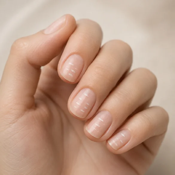
Leukonychia is the term for white discoloration of the nail plate, which can take the form of tiny spots, transverse or longitudinal bands, and sometimes cover the entire nail surface. Although for many people it is only an aesthetic problem, in dermatology it is an important diagnostic symptom, as it can result from both local damage to the nail apparatus and be a sign of systemic diseases.
Most often, leukonychia is the result of microtrauma to the nail matrix and is not associated with serious health disorders. However, there are situations in which the characteristic whitening of the nails requires thorough internal medicine, hepatological, or nephrological diagnostics.
Correct diagnosis of the type of leukonychia is crucial, because not every white lesion is located in the nail plate itself. Some lesions arise in the nail bed or are the result of an optical disturbance of light passing through the tissues. For this reason, dermatological evaluation includes not only the appearance of the nail, but also medical history, dermoscopy, and analysis of coexisting symptoms.
Leukonychia - what it is
Leukonychia (from Greek leukos – white, onyx – nail) means white discoloration of the nail caused by a disruption of its structure or changes occurring within the nail bed. It is not an independent disease, but a symptom with many possible causes.
In dermatology, the following are primarily distinguished:
- true leukonychia (leukonychia vera) – the changes originate in the nail matrix and are present in the plate itself,
- apparent leukonychia – the white color results from changes within the nail bed,
- pseudoleukonychia – the white discoloration is the result of surface changes, e.g., a fungal infection or external damage.
The mechanism of true leukonychia formation is related to abnormal keratinization of the cells forming the nail plate.
During this process:
- keratinocyte maturation is disrupted,
- microscopic cell nuclei remain in the plate (parakeratosis),
- tiny air-filled spaces appear between cells,
- light is scattered instead of penetrating through the transparent plate,
- the nail takes on a white color.
Since nails grow on average about 2–3 mm per month on the hands and about 1 mm per month on the feet, most changes move along with the growth of the plate and are cut off over time.
The mere presence of leukonychia does not indicate a serious disease. Clinical significance is determined primarily by the appearance of the changes, the number of affected nails, the patient's age, and coexisting systemic symptoms.
Leukonychia - white spots, stripes, completely white nails - types
Leukonychia can take several characteristic forms that differ in their mechanism of formation and diagnostic significance.
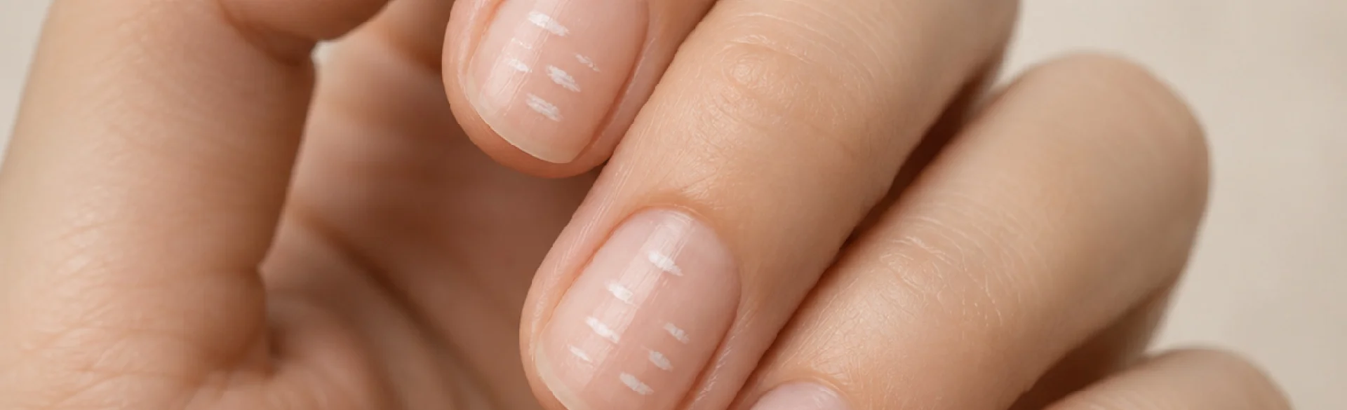
Leukonychia punctata (punctate)
It is the most common form.
It is characterized by:
- small white spots,
- single or numerous dots,
- gradual movement towards the free edge of the nail.
It is most often the result of minor injuries to the nail matrix.
Micro-injuries occur, among others, during:
- manicure,
- nail biting,
- intense tapping with nails,
- performing manual work.
This form is practically always benign.
Leukonychia striata (transverse)
It manifests as white transverse or longitudinal lines.
It may appear after:
- injuries,
- severe infections,
- chemotherapy,
- temporary inhibition of nail matrix activity.
A special type are Mees' lines and Muehrcke's lines, which have different clinical significance and help in the diagnosis of systemic diseases.
Partial leukonychia
It covers a significant part of the nail plate.
Changes may affect:
- half of the nail,
- its distal part,
- its proximal part.
It may occur in both local and systemic diseases.
Leukonychia totalis
The entire nail plate becomes milk-white.
It can be:
- congenital,
- familial,
- acquired.
Acquired leukonychia totalis is a rare symptom and requires in-depth diagnostics.
Apparent leukonychia
In this form, the nail plate itself remains normal.
The white coloration results from changes occurring under the nail.
The most well-known include:
- Terry’s nails,
- Lindsay’s nails,
- Muehrcke’s lines.
Their presence may indicate liver or kidney diseases, circulatory failure, or protein metabolism disorders.
Correct differentiation of the type of leukonychia often requires dermoscopic examination and assessment of how the lesion disappears upon compression of the plate.
Leukonychia - the calcium deficiency myth - what really causes it
One of the most widespread myths about nails is the belief that white spots indicate a calcium deficiency.
Modern dermatology clearly shows that in most people, such a connection does not exist.
The most common causes of leukonychia are:
- micro-injuries to the nail matrix,
- mechanical or hybrid manicure performed with damage to the nail fold,
- nail biting,
- frequent impacts to the plate,
- chronic mechanical irritation,
- contact with chemical substances,
- the use of aggressive solvents.
Less frequently, leukonychia may be associated with:
- zinc deficiency,
- severe protein deficiency,
- certain metabolic disorders,
- heavy metal poisoning,
- the effects of cytostatic drugs,
- genetic diseases.
Contrary to popular belief:
- calcium deficiency practically does not cause typical white spots on the nails,
- routine intake of calcium preparations does not eliminate leukonychia,
- supplementation without diagnosing the actual cause does not yield results.
If the lesions appear individually and move with nail growth, they usually do not require treatment. The most important thing is to reduce trauma and properly care for the nail apparatus.
Leukonychia - when white nails are a symptom of an internal disease
Although most cases of leukonychia are benign, certain types of changes can be a valuable sign of systemic diseases.
Particular attention should be paid to situations when:
- all nails are affected,
- whitening appears suddenly,
- leukonychia coexists with weakness, weight loss, or swelling,
- the changes do not move as the nail grows,
- other abnormalities of the skin, hair, or mucous membranes occur simultaneously.
White nails can accompany, among others:
- liver cirrhosis,
- chronic renal failure,
- nephrotic syndrome,
- severe hypoalbuminemia,
- heart failure,
- diabetes,
- gastrointestinal diseases with malabsorption disorders,
- autoimmune diseases,
- certain genetic syndromes.
Of particular diagnostic significance are:
- Terry’s nails – almost completely white with a narrow pink band at the free edge, often observed in liver cirrhosis, heart failure, and diabetes,
- Lindsay’s nails (half-and-half nails) – the upper part of the nail is white, and the distal part is brownish-red; characteristic of chronic kidney disease,
- Muehrcke’s lines – transverse white bands associated with decreased blood albumin concentration.
Diagnostics of leukonychia primarily include a thorough medical history, dermatological examination, and dermoscopic evaluation of the nails. In justified cases, the doctor may order laboratory tests, including complete blood count, liver and kidney function parameters, albumin concentration, zinc levels, and evaluation of metabolic status.
Most white spots on nails turn out to be completely harmless and disappear as the nail plate grows. However, if the changes are extensive, affect multiple nails, or are accompanied by systemic symptoms, they should be treated as a signal requiring professional diagnostics. In dermatology, the appearance of nails often provides valuable information about the health of the entire organism and can be one of the first visible symptoms of a developing disease.
