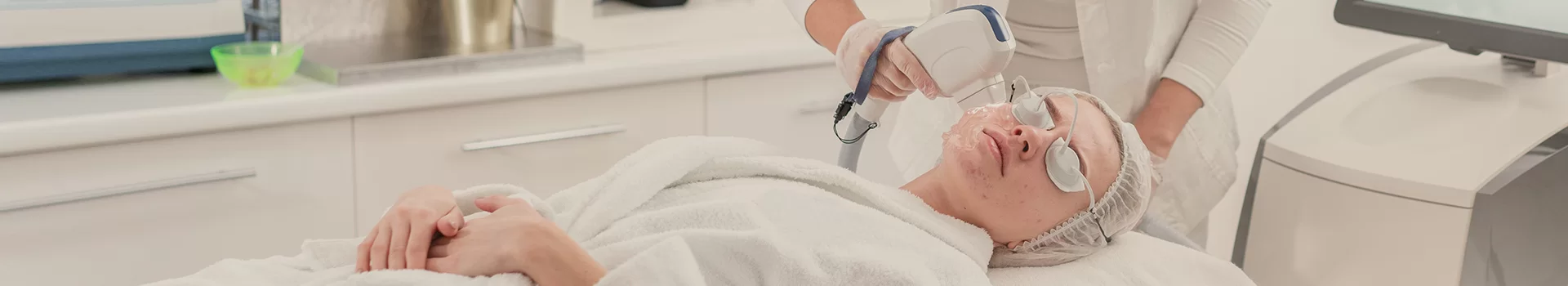
Although hemangiomas are usually not a health threat, they can be unsightly, especially when they are in visible areas of the body, such as the face or hands. In such cases, due to their impact on appearance, many people choose to have them removed to improve their psychological and aesthetic comfort. Laser removal is an ideal solution because it works precisely, removing the blemish without damaging surrounding tissues, ensuring a quick return to normal skin appearance without the need for traditional surgical methods. The procedure is safe and painless, and most importantly, it does not leave any scars behind.
WHAT ARE CAPILLARIES?
Hemangiomas are benign lumps that form as a result of excessive development of small blood vessels. They are considered tumor-like growths, but due to their non-malignant nature, they do not pose a threat to health. The occurrence of hemangiomas on the skin depends on the type of hemangioma. They most commonly appear on the upper half of the body, where the skin is thin and delicate, such as on the face, neck, décolletage, arms, hairy skin of the head, hands, fingers, torso, and less frequently on other parts of the body.
Hemangiomas are not large and usually are the size of a pinhead. They often have an intensely pink or ruby color, but sometimes they can be slightly bluish or brown, especially if they persist on the skin for a longer time. A distinct border can be observed between the hemangioma and healthy skin, although a smooth edge may not be present.
TYPES OF BIRTHMARKS
- Spider angiomas appear as numerous small red bumps branching out from a central point, resembling spider legs. They fade under pressure and are most commonly found on the face, upper chest, back, and arms. They usually affect women and children more often than men. They can occur as single lesions on the skin, but multiple lesions are more common. Spider angiomas do not cause pain or pose a health risk, but can be a significant cosmetic concern.
- Ruby angiomas (senile angiomas) are bright red in color and typically have a round, raised shape. They range in size from one to several millimeters and may look like soft, red balls of clay stuck to the skin. They can be found anywhere on the body. Ruby angiomas are benign growths that usually appear in adults over the age of thirty, with their numbers increasing with age. They do not pose any health risks, but they do not tend to spontaneously regress.
- Flat angiomas are non-raised red patches on the skin. They are most commonly seen in children, often appearing in infancy and tending to fade over time. If a flat angioma persists on the skin when the child is around 12 years old, it is likely to be a permanent change.
WHICH HEMANGIOMAS QUALIFY FOR LASER THERAPY?
Laser therapy is an effective and safe method for treating benign vascular skin lesions, but not every hemangioma requires or qualifies for the procedure. The type of lesion, its depth, extent, and clinical stability are of key importance. Qualification is always done individually, after specialist evaluation.
The following qualify for laser therapy:
- cherry angiomas (senile) – both single and multiple, regardless of location on the face or body
- spider angiomas – particularly visible lesions located on the face, neck, and décolletage
- small, superficial hemangiomas – red in color, clearly visible through the epidermis
- flat vascular lesions that do not show a tendency to spontaneously disappear
- diffuse vascular lesions and vascular erythema, requiring work on a larger skin area
- vascular lesions of an aesthetic nature, which do not pose a health threat but affect the skin's appearance and patient's comfort
The following do not qualify for laser therapy or require special caution:
- lesions of ambiguous nature, requiring prior diagnosis
- pigmented lesions or those suspicious of being oncological
- deep hemangiomas or those with a structure requiring different therapeutic management
- lesions in the phase of active inflammation
The final decision regarding the possibility of performing laser therapy is made after clinical evaluation, and if necessary, also after a dermatoscopic examination. This approach ensures the safety of the procedure and a predictable, aesthetic treatment outcome.
CONTRAINDICATIONS FOR LASER REMOVAL OF HEMANGIOMAS
Laser removal of hemangiomas is a safe procedure, provided that the patient is properly qualified. However, there are situations in which the procedure should not be performed or requires postponement. Each case is assessed individually during a consultation.
- suspected neoplastic change or lack of unequivocal dermatological qualification
- pigmented changes of uncertain nature in the treatment area
- active skin infections (bacterial, viral, fungal), including herpes
- skin inflammations, epidermal damage, fresh wounds, or abrasions
- skin diseases in an acute phase (e.g., atopic dermatitis, psoriasis)
- uncontrolled diabetes and other systemic diseases impairing the healing process
- blood coagulation disorders and the use of anticoagulants (requiring individual assessment)
- tendency to form hypertrophic scars and keloids
- autoimmune diseases in an active phase
- pregnancy and breastfeeding period (relative contraindication)
- recent suntan or planned intense UV exposure
- use of photosensitizing drugs and herbs
- recently completed retinoid therapy (isotretinoin)
In the presence of contraindications, the procedure may be postponed, the therapy method may be changed, or the patient may be referred for additional diagnostics. Such an approach constitutes a safety standard and allows for predictable, aesthetic treatment outcomes.