Congenital nevus
back to main page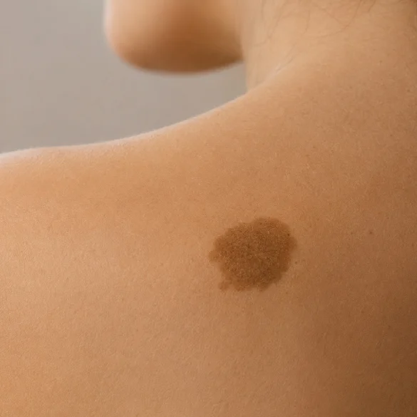
Congenital nevus (Latin: congenital melanocytic nevus, CMN) is a benign pigmented skin lesion present already at birth or appearing in the first weeks of life. It develops as a result of disturbances in the migration and differentiation of melanocytes, i.e., melanin-producing cells, during fetal life. Congenital nevi differ from each other in size, depth of skin infiltration, clinical appearance, and the risk of developing complications. Most remain benign lesions throughout life; however, large and giant nevi require regular dermatological check-ups due to the increased risk of developing melanoma and the possible coexistence of other developmental disorders. Modern dermoscopic diagnostics allows for a precise assessment of such lesions and proper planning of further management.
Congenital birthmark - what is it
A congenital nevus is a melanocytic hamartoma, which is a benign proliferation of melanocytes developing already during the fetal period. Melanocytes in congenital nevi are distributed not only in the epidermis and dermis, but often also deeper, in the subcutaneous tissue, around hair follicles, sebaceous glands, vessels, and nerves. It is this deeper location that distinguishes them from most acquired nevi.
It is estimated that congenital nevi occur in about 1% of newborns, however, the vast majority are small-sized lesions.
Characteristic features of a congenital nevus include:
- presence from birth or appearance in the first weeks of life,
- coloration from light brown to dark brown or almost black,
- a smooth, warty, or uneven surface,
- possible presence of dense hair,
- gradual growth proportional to the child's growth.
During puberty and in adulthood, the appearance of the nevus may undergo natural changes. The surface often becomes more uneven, hair appears, and the color may deepen or lighten locally. These changes do not necessarily indicate a neoplastic process, however, any new asymmetry, rapid growth, or ulceration requires a dermatological consultation.
Diagnosis is based primarily on clinical and dermoscopic examination. In the case of large lesions or diagnostic doubts, imaging studies or a histopathological examination after removal of the lesion are additionally performed.
Congenital birthmark - small, medium, giant - how do they differ
The most important factor influencing prognosis is the size of the nevus after reaching adulthood. In clinical practice, a classification taking into account the predicted diameter of the lesion is used.
Type of nevus | Size in adulthood | Characteristics |
|---|---|---|
Small | below 1.5 cm | Most common, low risk of complications |
Medium | 1.5-20 cm | Usually require periodic follow-up |
Large | 20-40 cm | Higher risk of melanoma, surgical treatment is often considered |
Giant | over 40 cm | High risk of dermatological and neurological complications |
Small congenital nevi
They constitute the vast majority of all cases. They usually remain benign lesions throughout life, and the risk of malignant transformation is very low. Dermatological control consists mainly of periodic dermoscopic evaluation.
Medium congenital nevi
They require more regular observation, especially when located in areas exposed to chronic mechanical irritation, such as the waist area, feet, hands, or scalp.
Large and giant congenital nevi
They cover extensive areas of skin and may occupy a significant part of the trunk, limbs, or head. Often, there are numerous smaller satellite nevi distributed around the main lesion.
In this group, there is an increased risk of:
- development of melanoma,
- neurocutaneous melanocytosis, i.e., the presence of melanocytes in the meninges,
- aesthetic problems,
- psychological disorders related to appearance.
Patients with large nevi usually remain under the care of a dermatologist, a plastic surgeon, and, if necessary, a neurologist.
Congenital nevus - is it more dangerous than an acquired one
Not every congenital nevus is associated with a greater risk than an acquired nevus. The risk depends primarily on its size, location, and clinical presentation.
Small congenital nevi show only a slightly higher risk of developing melanoma than common acquired nevi and in most cases remain completely benign.
Large and giant nevi are of much greater clinical significance. Within them, melanoma can develop:
- superficially,
- in deeper layers of the skin,
- in the subcutaneous tissue,
- much earlier than in the case of acquired nevi, sometimes even in childhood.
It is worth emphasizing that prophylactic removal of a nevus does not completely eliminate the risk of developing melanoma, as melanocytic cells may also remain in deeper tissues.
Particular attention should be paid to warning signs:
- rapid enlargement of the lesion,
- the appearance of irregular borders,
- significant asymmetry,
- color variation,
- bleeding,
- ulceration,
- chronic pain or itching.
Such symptoms require urgent dermatological consultation and often a biopsy or excision of the lesion.
Congenital birthmark - when to remove it
Not every congenital nevus requires surgical treatment. The decision is always based on an individual assessment of the risk of malignancy, the possibility of complete removal of the lesion, and the predicted aesthetic outcome.
The most common indications for removal include:
- suspected malignant transformation,
- ambiguous dermoscopic appearance,
- rapid changes in the appearance of the nevus,
- chronic mechanical irritation,
- frequent trauma,
- functional considerations,
- significant aesthetic defect or psychological burden.
The standard of treatment remains surgical excision of the lesion with a margin of healthy tissue and mandatory histopathological examination. In the case of large and giant nevi, treatment is often carried out in stages and may involve the use of tissue expanders or surgical reconstruction.
Laser therapy is not a recommended method for removing melanocytic nevi suspected of malignancy, as it does not allow for a full histopathological evaluation of the removed tissue. It can only be used in strictly selected cases and after prior qualification by a specialist.
Regardless of the treatment used, individuals with congenital nevi should remain under periodic dermatological follow-up. Regular digital dermoscopy enables the detection of even minor changes indicating malignant transformation at a very early stage, when treatment offers the best prognosis.

