Implant scar
back to main page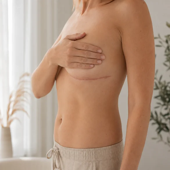
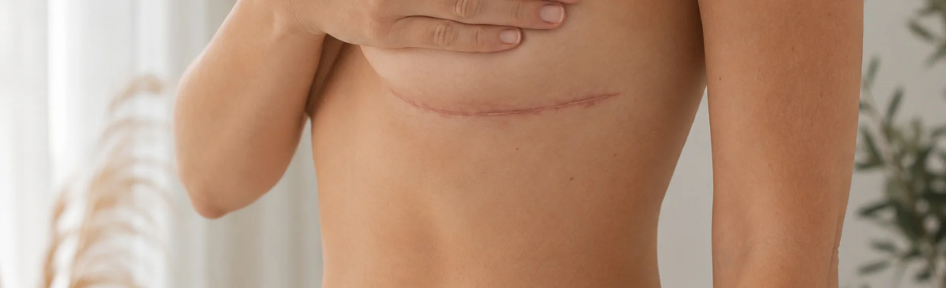
A breast implant scar is a natural consequence of a surgical skin incision through which the implant is inserted into the prepared pocket. Its location depends on the surgical approach used, while its appearance depends on the wound closure technique, tissue tension, the healing process, and individual biological predispositions. Initially, the scar may be pink, firm, slightly raised, and sensitive. It usually gradually fades, softens, and becomes flatter, but its full remodeling takes many months. A surgical scar should not be confused with capsular contracture, which develops deeper, around the implant. Sudden redness, worsening pain, swelling, discharge, or fever require urgent surgical evaluation.
Implant scar - where it runs depending on the incision
The placement of the scar depends on the surgical approach chosen by the plastic surgeon. The decision is made taking into account breast anatomy, the type and size of the implant, the surgical plan, skin quality, previous procedures, and the patient's preferences. There is no single incision that is optimal for everyone.
Most commonly used surgical approaches:
- Inframammary fold incision runs in the natural crease of the skin under the breast. It is currently one of the most frequently chosen approaches because it provides the surgeon with direct control over pocket dissection and implant positioning. The scar remains on the breast, but when properly planned, it is usually concealed by its lower pole. The position of the fold may change after the implant is inserted, which is why the incision site must be precisely marked before surgery.
- Periareolar incision most often runs along the lower border of the areola. The difference in color between the areola and the skin can partially mask the scar. This approach requires an adequate areolar diameter and involves cutting through part of the glandular tissue. It may affect nipple sensation and, depending on the extent of the surgery, the ability to breastfeed.
- Axillary incision is located in the natural fold of the armpit, so no scar remains on the breast itself. The implant is inserted through a longer surgical tunnel, often using an endoscope. The scar may be visible when raising the arm, and performing certain revision surgeries may require a different approach.
- Transumbilical incision, or the transumbilical approach, is rarely used and is not suitable for every type of implant or surgical plan. The scar is located within the navel, but control over pocket dissection is more limited.
If breast augmentation is combined with mastopexy, or breast lift, the extent of the scars is greater. They may run around the areola, vertically from the areola to the inframammary fold, and horizontally in the fold, forming an inverted T shape. The length of the scar in itself does not determine its final quality.
Scar after implants - how long does it take to mature
Wound healing and scar maturation are biological processes involving overlapping phases. Immediately after surgery, hemostasis occurs, during which blood platelets and the coagulation system limit bleeding. Then, a controlled inflammatory phase develops, necessary for wound cleansing and initiating tissue repair.
In the proliferative phase, fibroblasts synthesize extracellular matrix components, primarily type III collagen. New blood vessels are formed, which is why a fresh scar is usually pink or red. At the same time, myofibroblasts cause wound contraction.
The approximate course of maturation is as follows:
- first 1-2 weeks: wound closure, swelling, and possible bruising;
- 2-8 weeks: increase in fibroblast activity, vascularity, and the amount of collagen; the scar may become firmer and more visible;
- 2-6 months: gradual reorganization of collagen fibers, reduction in vascularity, and tissue softening;
- 6-18 months: further remodeling, fading, and flattening of the scar;
- up to 24 months: in some patients, subtle changes in scar color, elasticity, and width may still occur.
During remodeling, type III collagen is partially replaced by stronger type I collagen, and its fibers align according to the acting mechanical forces. A scar never regains the full structure and strength of undamaged skin.
The rate of maturation depends, among other factors, on genetics, age, skin phototype, tobacco smoking, diabetes, malnutrition, infection, wound dehiscence, and wound tension. Assessment of the final appearance of the scar should not be performed after just a few weeks, as this is the period of its greatest metabolic activity.
Scar after implants - why it is sometimes hard and red
Transient firmness and redness are most often part of normal healing. A fresh scar contains an increased number of small vessels, inflammatory cells, fibroblasts, and immature collagen fibers. The regulation of these processes is governed by, among others, transforming growth factor beta, interleukins, and mechanical tissue tension.
A scar requires evaluation when its features are excessive, worsen, or persist longer than expected based on the healing stage. The following should be considered:
- hypertrophic scar, which is red, raised, firm, sometimes itchy or painful, but remains within the boundaries of the original incision;
- keloid, which grows beyond the boundaries of the wound and tends to recur; its formation is favored by individual and familial predispositions;
- scar widening, associated with skin tension, tissue movement, delayed healing, or separation of the wound edges;
- suture reaction, which may cause focal thickening, redness, or a small tract with discharge;
- infection, especially when increasing pain, localized warmth, swelling, pus, foul odor, fever, or wound dehiscence occur;
- post-inflammatory hyperpigmentation, more common in higher phototypes and after exposure of a fresh scar to ultraviolet radiation.
Hardness limited to the skin differs from hardening of the entire breast. Increasing breast firmness, a change in its shape, implant elevation, pain, or asymmetry may indicate capsular contracture. This is a complication affecting the tissue surrounding the implant, rather than the superficial skin scar.
Any sudden breast enlargement, late swelling, presence of fluid, or a lump in the breast or axilla requires consultation with a plastic surgeon. Such symptoms should not be treated with aesthetic procedures without first determining their cause.
Implant scar - how to take care of it
Management should take into account the healing stage and the surgeon's recommendations. Until the wound is fully closed, massage, silicone products, or procedures that disrupt the epidermis should not be used without the surgeon's consent. The dressing and wound must be cared for according to the provided instructions, without picking scabs or self-administering topical antibiotics.
After complete epidermal healing, the following are most commonly considered:
- silicone gel or sheets, which reduce transepidermal water loss, improve hydration of the stratum corneum, and can reduce hardness, redness, and the risk of scar hypertrophy;
- wound tension reduction using appropriately selected tapes, if recommended by the surgeon;
- scar massage, introduced only after proper healing is confirmed; it can improve tissue mobility and elasticity, although the protocol should be individually tailored;
- SPF 50+ photoprotection and shielding the scar from the sun, especially during the first year, to reduce the permanence of hyperpigmentation;
- avoiding smoking and nicotine, which impair microcirculation and increase the risk of healing complications;
- following recommendations regarding the post-operative bra, physical activity, and heavy lifting.
When a scar becomes hypertrophic, the doctor may use corticosteroid injections, sometimes combined with 5-fluorouracil, laser therapy, or combination treatment. Surgical scar revision is reserved for selected cases and usually requires the simultaneous reduction of factors responsible for its abnormal formation.
The offer of Ambasada Urody Clinic & Spa also includes treatments intended for breast augmentation scars. Following medical qualification and confirmation of stable healing, the following may be considered:
- fractional CO₂ laser and other ablative fractional lasers, which create microscopic columns of ablation and coagulation, initiating collagen remodeling;
- non-ablative fractional laser, which heats the dermis without epidermal ablation and is typically associated with a shorter recovery time;
- OScar hybrid laser, combining ablative and non-ablative effects to target the surface and deeper structure of the scar;
- microneedle radiofrequency, which delivers thermal energy to the dermis via microneedles and can improve tissue elasticity and structure;
- platelet-rich plasma, mesotherapy, and LED therapy as supportive methods for regeneration in appropriately selected combined protocols.
The type of device, parameters, and the starting date of therapy require individual determination. A fresh, unhealed, or infected scar is not subjected to resurfacing procedures. No method removes a scar completely, but properly planned treatment can reduce its redness, thickness, hardness, and visibility.

