Blemish
back to main page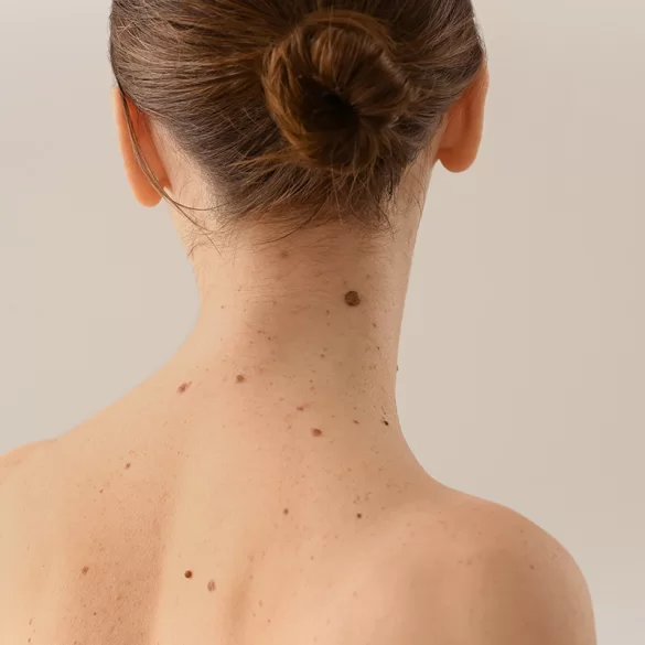

A mole is a limited skin alteration resulting from the local accumulation of melanocytes (pigment cells) or other skin structures. Moles can be congenital or acquired and differ in size, color, shape, and location. In most cases, they are benign changes, but some require regular dermatological observation due to the potential risk of cancerous transformation. The assessment of moles is based on clinical examination, dermatoscopy, and — in justified cases — histopathological analysis. Modern dermatology and skin surgery have effective, safe methods for diagnosing and removing moles, tailored to their type, location, and medical indications.
Birthmark – what it looks like
The appearance of a mole is varied and depends on its histological type and the depth of pigment cell placement. The most commonly encountered are pigmented moles, but there are also epidermal, vascular, and connective tissue moles.
Typical features of moles:
- color: from light beige, through brown, to almost black; less commonly pink or reddish,
- shape: round or oval,
- surface: smooth, warty, or slightly elevated,
- size: from a few millimeters to several centimeters,
- borders: usually clear and regular.
In clinical practice, the ABCDE rule is used, which helps in the assessment of pigmented moles:
- A (asymmetry) – asymmetry of shape,
- B (border) – irregular, jagged edges,
- C (color) – uneven coloration,
- D (diameter) – diameter > 6 mm,
- E (evolution) – change in appearance over time.
The appearance of one or more of the above characteristics requires urgent dermatological consultation.
Birthmark - Is it Dangerous
The vast majority of moles are benign and do not pose a health threat. However, there are changes that may undergo malignant transformation, particularly towards melanoma.
Factors increasing the risk of mole malignancy:
- intense and chronic exposure to UV radiation,
- numerous sunburns during childhood,
- light skin phototype (I–II),
- a large number of pigmented moles (>50),
- presence of atypical moles,
- positive family history of melanoma.
Warning symptoms include:
- rapid growth of the lesion,
- change in color or shape,
- bleeding, oozing, itching, or pain,
- the onset of inflammation around the mole.
Any suspicion of a change in the nature of a mole is an indication for specialist diagnostics, and if necessary, for its removal.
Birthmark – treatment
The management of a mole depends on clinical and dermatoscopic evaluation. For benign moles, regular dermatological check-ups are usually sufficient, especially for patients in high-risk groups.
Possible management strategies:
- periodic observation (manual or digital dermoscopy),
- prophylactic removal of a mole exposed to mechanical injuries,
- diagnostic removal in case of an ambiguous appearance,
- surgical treatment with histopathological examination.
Self-removal of moles or using "burning" preparations is not recommended because it can:
- delay cancer diagnosis,
- complicate histopathological assessment,
- increase the risk of complications and scarring.
The therapeutic decision should always be made by a physician, based on current guidelines and the clinical condition of the patient.
Birthmark – Removal Methods
The choice of mole removal method depends on its type, depth, location, and medical indications.
1. Surgical excision of the mole
- preferred method when malignancy is suspected,
- allows for a complete histopathological examination,
- performed under local anesthesia,
- leaves a scar depending on technique and location.
2. Shave excision
- used for selected benign moles,
- no need for stitching,
- limited possibilities for histopathological evaluation.
3. Laser therapy
- used exclusively for benign moles,
- no material for histopathological examination,
- an aesthetic method requiring strict qualification.
4. Electrosurgery / radiofrequency
- precise removal of superficial lesions,
- short healing time,
- only after excluding atypical features.
The choice of technique must be preceded by thorough diagnostics, and the primary goal is the oncological safety of the patient.
Mole removal treatments
In clinical conditions, mole removal procedures are performed in accordance with the principles of asepsis and medical safety. The procedure includes dermatological qualification, selection of the method, and—if indicated—preservation of material for histopathological examination.
Standard procedure:
- dermatological consultation and dermatoscopy,
- qualification for a specific removal method,
- procedure under local anesthesia,
- post-procedure instructions (UV protection, wound care),
- post-procedure check-up and discussion of histopathology results.
Mole removal procedures include surgical excision of lesions, electro-surgical methods, and laser methods, each time following specialist medical qualification and adhering to current dermatology and skin surgery standards.
