Cherry angioma
back to main page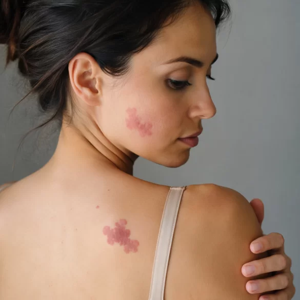
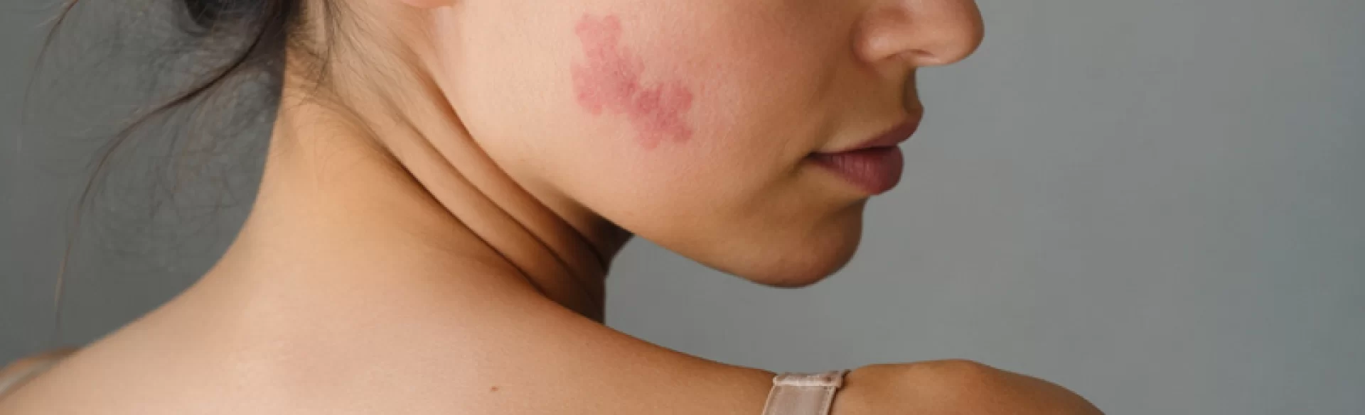
Cherry angioma (senile angioma, angioma rubinum) is a benign skin lesion of vascular origin, resulting from the proliferation (multiplication) of small blood vessels within the dermis. These lesions occur most frequently in adults, and their number increases with age. Cherry angioma is benign and does not show a tendency for neoplastic transformation; however, for aesthetic or diagnostic reasons, it often becomes the subject of dermatological consultation. The clinical presentation is characterized by typical small red dots or papules, which may occur singly or in multiples, mainly on the trunk.
Cherry angioma – what it looks like
A cherry angioma takes the form of small, well-demarcated skin lesions with a diameter of 1 to several millimeters, although in some cases they can reach larger sizes. Characteristic morphological features include:
- intensely red, ruby, or cherry coloration,
- a smooth, dome-shaped surface,
- absence of inflammatory symptoms in the vicinity of the lesion,
- a tendency to be slightly elevated above the skin surface.
These lesions are most commonly located on:
- the trunk (chest, abdomen, back),
- the arms,
- less frequently on the face and lower limbs.
In a dermatoscopic examination, characteristic vascular structures are visible - so-called “blood lakes” (lacunae), corresponding to dilated vascular spaces filled with erythrocytes. A cherry angioma usually does not cause pain or itching; however, it may bleed in the case of mechanical trauma, which results from its rich vascularization.
Cherry angioma – is it dangerous
Cherry angioma is a completely benign lesion and does not pose an oncological risk. It does not show the potential for malignancy, which distinguishes it from some other pigmented or vascular skin lesions. Its presence is also not associated with systemic disorders.
In clinical practice, however, it is important to differentiate cherry angiomas from other skin lesions, in particular:
- amelanotic melanoma (pigment-free),
- cavernous hemangiomas,
- pyogenic granuloma,
- telangiectasias.
Diagnostic concern may be raised by lesions with an atypical course, such as:
- rapid growth,
- irregular shape,
- change in color,
- tendency to spontaneous bleeding.
In such cases, a dermatological consultation and a possible dermoscopic or histopathological examination are recommended. It is worth emphasizing that the number of cherry angiomas may increase with age, and their appearance is sometimes associated with genetic predisposition, hormonal changes, or skin aging processes.
Cherry angioma – removal
Removal of cherry angiomas is performed mainly for aesthetic indications or in the case of diagnostic doubts. Modern aesthetic medicine and dermatology offer a range of effective and safe therapeutic methods, the choice of which depends on the size, location, and number of lesions.
The most commonly used methods include:
- vascular laser therapy – the use of selective photothermolysis, a phenomenon involving selective damage to blood vessels by laser light (e.g., dye laser, Nd:YAG); it leads to the closure and gradual disappearance of the lesion,
- electrocoagulation – a method involving the coagulation of proteins using an electric current, which causes the destruction of the lesion,
- cryotherapy – the use of low temperature (liquid nitrogen) to destroy pathological vessels,
- radiosurgery – precise removal of the lesion using high-frequency radio waves.
In clinical settings, such as Ambasada Urody Clinic & SPA, modern laser technologies are of particular importance, as they enable:
- high selectivity of action,
- minimization of damage to surrounding tissues,
- short recovery time,
- a very good aesthetic effect.
After the procedure, transient erythema or slight swelling may occur, which resolve spontaneously within a few days. In most cases, one therapeutic session is sufficient, although staged treatment is recommended for numerous lesions.

