Cherry angiomas
back to main page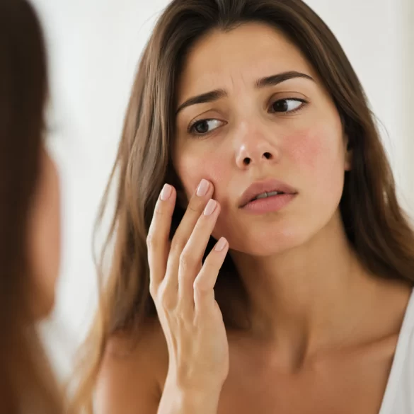
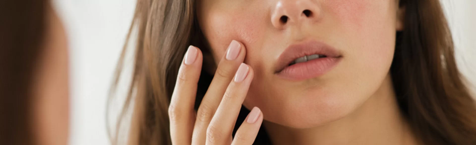
Cherry angiomas, also referred to as senile angiomas (angioma senile), are benign proliferations of the skin's blood vessels, manifesting as small, intensely red, burgundy, or ruby papules. They are most commonly located on the trunk, less frequently on the limbs and face. Their diameter usually ranges from 1 to several millimeters, and the number of lesions increases with age. These lesions are benign in nature and do not exhibit potential for malignant transformation. In dermatological practice, they are a frequent clinical finding, requiring differentiation from other vascular and pigmented lesions.
Cherry angiomas – causes
The etiopathogenesis of cherry angiomas is primarily associated with skin aging processes and angiogenesis disorders, i.e., the formation of new blood vessels. Local proliferation of endothelial cells and dilation of existing capillaries within the dermis occur.
The most important predisposing factors include:
- age – the lesions most often appear after the age of 30, and their number increases over time,
- genetic factors – familial occurrence of the lesions is observed,
- hormonal factors – the lesions may intensify during periods of hormonal imbalances,
- oxidative stress – cellular damage caused by free radicals,
- environmental factors – chronic exposure to UV radiation may exacerbate their development.
At the molecular level, the overexpression of proangiogenic factors is significant, including VEGF (vascular endothelial growth factor), which stimulates the proliferation of endothelial cells and the formation of new vascular structures.
It is worth noting that the sudden appearance of numerous cherry angiomas in a short period (the so-called Leser–Trélat sign) may coexist with neoplastic processes, particularly within the gastrointestinal tract. In such cases, in-depth internal medicine and oncological diagnostics are recommended.
Cherry angiomas – diagnostics and differentiation
Diagnosis of ruby lesions is primarily based on clinical presentation and dermoscopic examination. In dermoscopy, characteristic, homogeneous vascular structures of red or dark red coloration are visible, often referred to as “blood lakes”.
Differential diagnosis primarily includes:
- port-wine stains and cavernous hemangiomas,
- telangiectasias (dilated capillaries),
- pyogenic granulomas (pyogenic granuloma) – rapidly growing lesions, prone to bleeding,
- amelanotic melanoma (amelanotic melanoma) – rare, but requiring exclusion,
- seborrheic keratoses with vascular coloration.
In cases of doubt, the following are used:
- digital dermoscopy,
- videodermoscopy,
- histopathological examination (after removal of the lesion).
Proper differentiation is crucial because some lesions mimicking cherry angiomas may be neoplastic in nature.
Cherry angiomas – removal
Cherry angiomas do not require treatment for medical reasons; however, their removal is a standard dermatological and aesthetic procedure. Indications mainly include aesthetic considerations, a tendency to bleed after injury, chronic mechanical irritation, and the need for diagnostic verification.
Modern methods of removing cherry angiomas are based on the selective destruction of pathological blood vessels while sparing the surrounding tissues. Selecting technology appropriate to the depth and diameter of the lesion is of key importance.
The most commonly used methods include:
1. Vascular laser therapy
It utilizes the phenomenon of selective photothermolysis – light energy (e.g., Nd:YAG, KTP laser) absorbed by hemoglobin leads to coagulation and closure of the vessel.
Advantages: high precision, minimal risk of scarring, very good aesthetic results.
2. IPL (Intense Pulsed Light) systems
A broadband light source used mainly for numerous, small lesions.
Advantages: the possibility of treating multiple lesions simultaneously.
3. Electrocoagulation
A method involving the thermal destruction of the lesion using high-frequency current.
Advantages: fast and effective procedure, wide availability.
4. Radiosurgery
A technique using radio waves for the precise removal of skin lesions.
Advantages: minimal tissue damage, very good healing.
5. Cryotherapy
The application of liquid nitrogen leads to the destruction of the lesion by freezing it.
Limitations: less control over the depth of action, risk of discoloration.
The selection of the method depends on:
- size and number of lesions,
- anatomical location,
- skin phototype,
- expected aesthetic effect.
Possible post-treatment reactions:
- transient erythema and swelling,
- small scabs,
- rarely post-inflammatory hyperpigmentation.
In clinical practice, laser and radiosurgical technologies are of particular importance, as they ensure the highest effectiveness and procedural safety.
Cherry angiomas – prognosis and prevention
Cherry angiomas are permanent and do not undergo spontaneous regression. After their removal, it is possible for new lesions to appear in other locations, which results from individual predisposition and skin aging processes.
The prognosis is very good – the lesions:
- are not malignant,
- do not cause systemic complications,
- do not affect the functioning of the body.
Preventive measures have limited effectiveness, however, they may support the condition of blood vessels:
- photoprotection – limiting the impact of UV radiation,
- antioxidant skincare – the use of substances that neutralize free radicals,
- healthy lifestyle – a diet rich in antioxidants, physical activity.
In clinical practice, regular observation of skin lesions and dermatological consultation in the event of their sudden appearance or a change in character are of key importance.