Dysplastic nevus
back to main page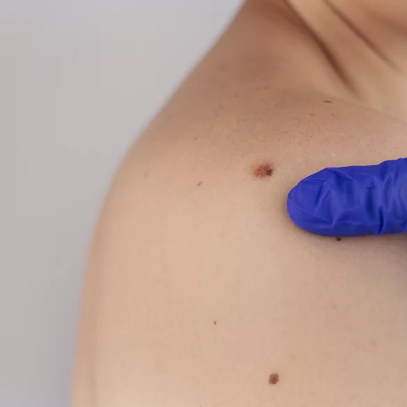
A dysplastic nevus (atypical mole) is a particular type of pigmented lesion that clinically and histopathologically exhibits characteristics that are intermediate between a typical melanocytic nevus and early skin cancer changes. It is characterized by an unusual appearance, irregular outline, heterogeneous coloration, and often larger size compared to classic benign moles. The presence of dysplastic nevi does not automatically indicate cancer, but it has significant diagnostic and prognostic importance because it is associated with an increased risk of developing melanoma. Therefore, they require special oncological vigilance, regular dermoscopic monitoring, and—in selected cases—surgical treatment.
Dysplastic nevus – what does it mean
A dysplastic nevus is a skin lesion resulting from abnormal proliferation of melanocytes – the cells that produce melanin. Unlike benign nevi, the melanocytes in dysplastic nevi exhibit architectural disturbances and features of cellular atypia, which may be visible in histopathological examination.
Clinical features of dysplastic nevi:
- asymmetry of shape,
- irregular, "ragged" edges,
- uneven coloration (ranging from light brown to black, sometimes with a hint of red),
- diameter often >5–6 mm,
- gradual change in appearance over time.
A dysplastic nevus can:
- occur singly,
- appear in large numbers (dysplastic nevus syndrome),
- develop on both sun-exposed and non-sun-exposed skin.
The clinical significance of such nevi lies in their being a marker of increased melanoma risk, especially in individuals with:
- fair skin phototype,
- numerous pigmented nevi,
- a positive family history of melanoma,
- a history of intense UV exposure.
Dysplastic nevus and melanoma
The relationship between a dysplastic nevus and melanoma is one of risk, not disease identity. This means that a dysplastic nevus is not melanoma, but it can:
- increase the overall risk of developing melanoma,
- rarely be the site of malignant transformation.
Epidemiological studies show that individuals with numerous dysplastic nevi have even several times higher risk of developing melanoma compared to the general population.
Key differences:
- dysplastic nevus → a benign lesion with atypical features,
- melanoma → malignant melanocytic tumor with metastatic potential.
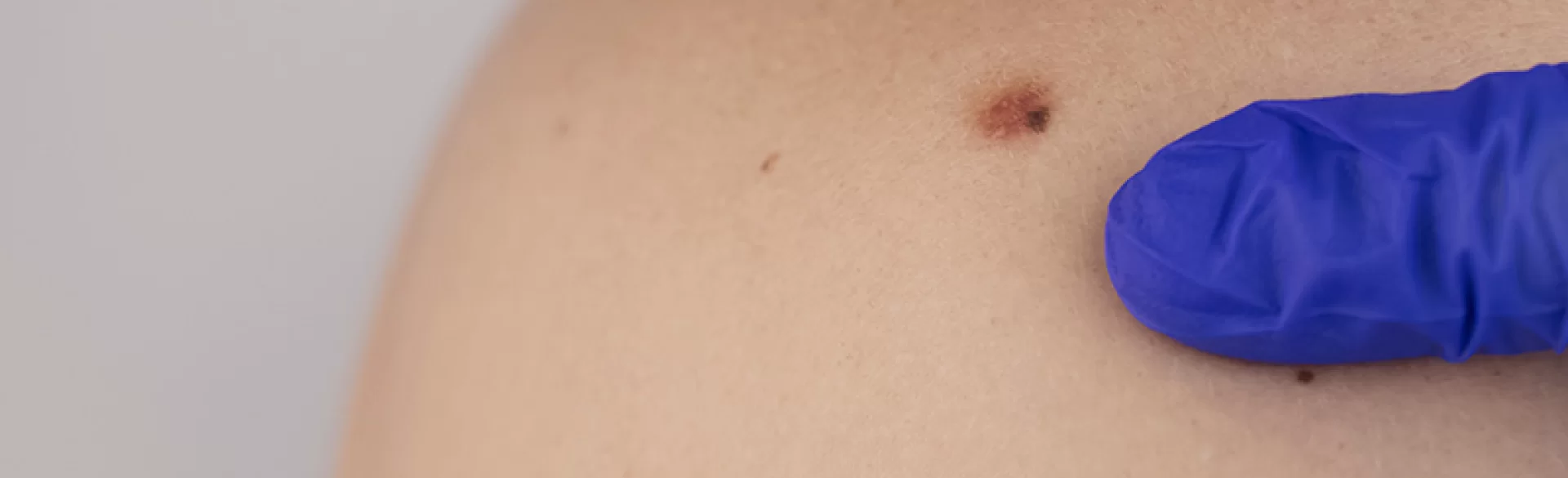
Warning signs (ABCDE) that may suggest lesion progression:
- A – increasing asymmetry,
- B – increasing irregularity of borders,
- C – changes in color or appearance of black, gray, red,
- D – dynamic increase in diameter,
- E – evolution (itching, bleeding, ulceration).
In case of suspected malignant transformation, the only appropriate course of action is urgent diagnosis and surgical treatment.
Dysplastic nevus – removal
The decision to remove a dysplastic nevus is based on clinical assessment, dermatoscopic evaluation, and the patient's history. Not every dysplastic nevus requires prophylactic excision, but there are clearly defined medical indications.
Indications for removal:
- features of high-grade atypia in dermatoscopy,
- dynamic changes in the appearance of the nevus,
- inability to perform regular observation,
- positive family history of melanoma,
- ambiguous diagnostic appearance.
The gold standard of management is:
- surgical excision of the nevus in its entirety,
- with preservation of a margin of healthy skin,
- with mandatory histopathological examination.
Destructive methods (laser, electrocoagulation, cryotherapy) are not recommended for suspicious or dysplastic nevi because:
- they preclude histopathological evaluation,
- they may mask malignant changes,
- they increase the risk of delay in melanoma diagnosis.
Treatments for dysplastic nevi
The therapeutic approach for dysplastic nevi is strictly medical and oncological, rather than aesthetic. The priority is the safety of the patient and a thorough diagnosis of the lesion.
Procedures used in clinical practice:
- surgical excision of the nevus with a scalpel,
- precise wound stitching with attention to scar aesthetics,
- histopathological analysis of the removed lesion,
- further management depending on the test results.
Supplementary clinical actions:
- regular dermatoscopy of the entire body,
- photographic documentation of changes,
- patient education in skin self-examination,
- individual dermatological observation plan.
For patients with numerous dysplastic nevi, treatment involves long-term dermatological-oncological care, rather than a one-time intervention.

