Fascia
back to main page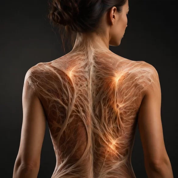

Fascia (Latin: fascia) is an extensive, three-dimensional system of connective tissue that penetrates and integrates all the body's structures – muscles, internal organs, vessels and the nervous system. It forms a continuous network with supportive, protective and communicative functions, participating in the transmission of mechanical forces and sensory stimuli. Contemporary research indicates that fascia not only serves as a 'wrapping' of tissues, but actively influences the biomechanics of movement, proprioception (deep sensation) and regenerative processes. Disorders of its structure and function can lead to restricted mobility, chronic pain and organ dysfunction.
Fascia – types
Fascia exhibits a varied structure and function depending on the anatomical location. In the classical view, three basic layers of fascia are distinguished:
1. Superficial fascia (fascia superficialis)
Located directly under the skin, it contains adipose tissue, blood vessels and nerve endings. It is responsible for:
- cushioning mechanical trauma,
- thermal insulation,
- allowing the skin to slide over deeper structures.
2. Deep fascia (fascia profunda)
It surrounds muscles, muscle groups and organ structures, forming intermuscular septa. Its functions include:
- stabilization of anatomical structures,
- transmission of mechanical tension,
- organization of muscle activity in functional chains.
3. Visceral fascia (fascia visceralis)
It surrounds internal organs, allowing their physiological mobility relative to one another. It participates in:
- maintaining the proper position of the organs,
- transmission of tensions between the abdominal cavity and the thoracic cavity,
- regulation of organ functions through mechanisms of mechanotransduction (the transmission of mechanical stimuli at the cellular level).
From a clinical point of view, fascia is considered a single continuous system in which a disturbance in one area may manifest as symptoms in distant parts of the body.
Fascia – role in stabilization and movement
The fascia exhibits varied structure and function depending on anatomical location. In the classical view, three basic layers of fascia are distinguished:
1. Superficial fascia (fascia superficialis)
Located directly beneath the skin, it contains adipose tissue, blood vessels, and nerve endings. It is responsible for:
- cushioning mechanical injuries,
- thermal insulation,
- allowing the skin to glide relative to deeper structures.
2. Deep fascia (fascia profunda)
Surrounds muscles, muscle groups, and organ structures, forming intermuscular septa. Its functions include:
- stabilization of anatomical structures,
- transmission of mechanical tensions,
- organizing muscle work into functional chains.
3. Visceral fascia (fascia visceralis)
Surrounds internal organs, allowing their physiological mobility relative to each other. It participates in:
- maintaining the proper position of organs,
- transmission of tensions between the abdominal and thoracic cavities,
- regulation of organ functions through mechanotransduction mechanisms (the transmission of mechanical stimuli at the cellular level).
From a clinical perspective, the fascia is regarded as a single continuous system in which a disturbance in one area can manifest as symptoms in distant parts of the body.
Fascia, pain, and muscle dysfunctions
The fascia is a densely innervated structure containing numerous pain receptors (nociceptors) and mechanoreceptors. For this reason it constitutes an important source of pain, particularly in cases of chronic pain syndromes.
The most common fascial disorders include:
- fascial restrictions – limitations of tissue mobility,
- trigger points (trigger points) – localized areas of increased tension,
- tissue hydration disorders – decreased water content and impaired sliding of fascial layers.
Symptoms of fascial dysfunction may include:
- pain that is diffuse or referred,
- a feeling of stiffness,
- reduced range of motion,
- impaired muscular coordination.
Contemporary concepts, such as the tensegrity model (balance of tension), emphasize that local fascial tensions influence the body's global biomechanics. This means that pain in one area can result from dysfunction in a completely different location.
Fascia and urogynecological physiotherapy
In urogynaecological physiotherapy the fascia plays a fundamental role in the functioning of the pelvic floor structures and the pelvic organs. The pelvic fascia forms a complex support system for:
- the urinary bladder,
- the uterus,
- the rectum.
Altered tension of the fascia may lead to:
- urinary incontinence,
- pelvic organ prolapse,
- chronic pelvic pain,
- dyspareunia (pain during intercourse).
The fascia's connections with the following structures are of particular importance:
- the diaphragm,
- the abdominal muscles,
- spinal structures.
Abnormal breathing biomechanics or increased intra-abdominal pressure can lead to overload of the pelvic fascia and deterioration of its supportive function.
Various approaches are used in therapy to normalize fascial tension, including:
- manual fascial therapy,
- myofascial release techniques,
- breathing and stabilization exercises,
- re-education of movement patterns.
Therapy may be supplemented by modern methods that support tissue regeneration, such as:
- radiofrequency microneedling – stimulating collagen remodeling,
- laser therapies – improving tissue elasticity and blood supply,
- procedures using platelet-rich plasma (PRP) – supporting regenerative processes.
An integrated approach to the fascia in urogynaecological physiotherapy enables effective treatment of many chronic conditions and the restoration of normal function of pelvic structures.