Halo nevus
back to main page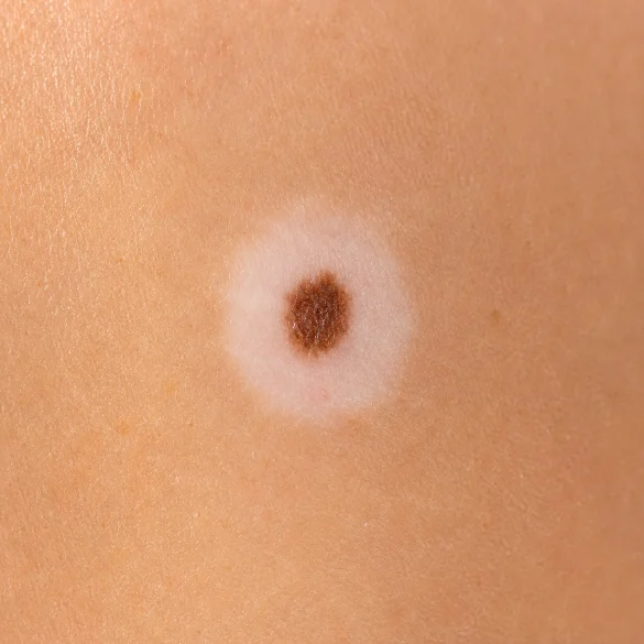
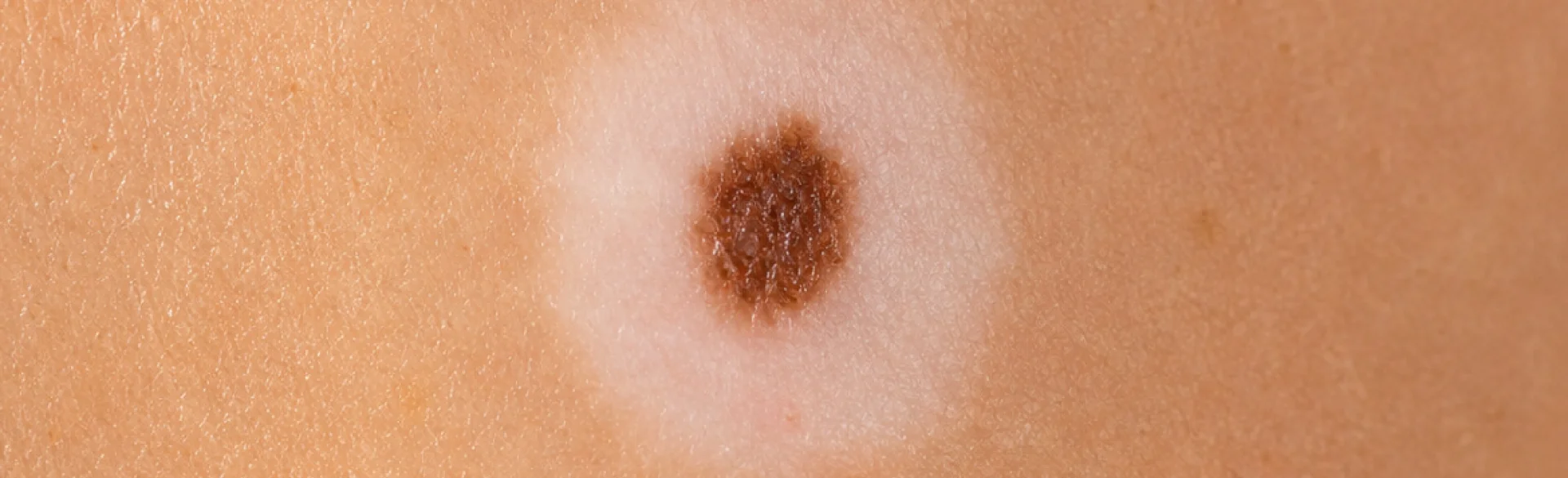
Halo nevus (Latin: naevus halo), also referred to as Sutton's nevus, is a particular type of melanocytic nevus surrounded by a characteristic, depigmented border of skin. This phenomenon results from an immune system reaction directed against melanocytes, i.e., cells that produce melanin. Although its appearance often causes concern in patients and may suggest skin cancer, in most cases, the halo nevus is benign. At the same time, any newly appearing white border around a nevus requires dermatological evaluation, as a similar clinical presentation can also occur in the course of regressing melanoma. In diagnostics, dermoscopic examination, analysis of the lesion's history, and evaluation of other skin nevi are of key importance.
Halo nevus - what is it
A halo nevus is an acquired melanocytic nevus around which a round or oval zone of skin depigmentation, devoid of melanin, develops. The mechanism of the lesion's formation is associated with the activation of T lymphocytes, which recognize melanocytes as cells requiring elimination. As a result, there is a gradual destruction of both the pigment cells within the nevus and those located in the surrounding epidermis.
Most commonly, the lesions appear in:
- children and adolescents,
- young adults,
- individuals with a predisposition to autoimmune diseases,
- patients with vitiligo.
A halo nevus can develop around any type of benign melanocytic nevus, but it most commonly affects acquired nevi located on the trunk, especially on the back. In some patients, a single lesion occurs, while in others, multiple halo nevi are observed, appearing simultaneously or gradually.
The lesion usually goes through several stages of development:
- the appearance of a white border,
- gradual decrease in the pigmentation of the nevus,
- complete disappearance of the nevus,
- slow repigmentation of the surrounding skin, which may take many years or not occur at all.
This process is completely natural and is a consequence of the body's immune response.
Halo nevus - what it looks like
The most characteristic symptom is a symmetrical, clearly demarcated white border surrounding the nevus. Its width is most often from a few to several millimeters, although it may be larger.
Typical clinical features include:
- a centrally located nevus with a regular shape,
- even coloration of the nevus,
- a round or oval zone of depigmentation,
- clear boundaries between the depigmented and normal skin,
- absence of features of inflammation.
In the following months or years, the nevus gradually:
- lightens,
- decreases in volume,
- may completely disappear.
After its resolution, only a white spot remains, which over time often regains the natural skin color.
During a dermoscopic examination, the dermatologist evaluates, among other things:
- symmetry of the lesion,
- the structure of the pigment network,
- pigment distribution,
- the presence of atypical vessels,
- features suggesting melanoma.
Dermoscopic examination allows distinguishing a typical halo nevus from lesions requiring removal and histopathological examination.
Halo nevus - where does the white border come from
The formation of the characteristic white border is the result of a local autoimmune reaction directed against melanocytes.
The most important role is played by:
- CD8+ T lymphocytes,
- pro-inflammatory cytokines,
- antibodies against melanocyte antigens.
Immune cells destroy melanocytes both within the nevus and in the surrounding skin. As a result, there is a loss of melanin production and depigmentation appears.
This process resembles the mechanism observed in acquired vitiligo, which is why both conditions often co-exist. In patients with multiple halo nevi, the risk of co-occurring vitiligo is higher than in the general population.
Factors contributing to the formation of the lesions include:
- genetic predisposition,
- autoimmune diseases,
- vitiligo,
- an intense immune response of the body.
However, halo nevus has not been shown to be caused by mechanical trauma, infection, or exposure to UV radiation, although these factors may influence the manifestation of lesions in predisposed individuals.
Halo nevus and Sutton's nevus - are they the same
Yes. The halo nevus and Sutton's nevus mean exactly the same disease entity.
The name “Sutton's nevus” comes from the American dermatologist Richard Lightburn Sutton, who described this lesion in detail in 1916. Currently, both terms function in parallel in medical literature:
- halo nevus (halo nevus) – a descriptive name referring to the white border,
- Sutton's nevus (Sutton nevus) – an eponymous name.
Both terms are fully equivalent and used interchangeably.
However, a classic halo nevus must be distinguished from:
- melanoma with features of regression,
- post-inflammatory depigmentation around skin lesions,
- vitiligo involving the area of the nevus,
- rare melanocytic lesions with an atypical course.
For this reason, the appearance of a white border around a nevus in an adult, especially after the age of 40, always requires thorough dermatological diagnostics.
Halo nevus - does the white border mean the mole is disappearing
In most cases, the answer is yes, although this process is gradual and can last from a few months to even several years.
The typical evolution of the lesion includes:
Stage | Clinical changes |
|---|---|
I | Appearance of a white border around the nevus |
II | Gradual fading of the nevus |
III | Complete disappearance of the nevus |
IV | Slow return of pigment within the depigmented skin |
Not every halo nevus disappears completely. In some patients, the central lesion persists for many years, despite the presence of the depigmented border.
Although a classic halo nevus is a benign lesion, a dermatological consultation is necessary when:
- a white border appears suddenly around a nevus with an irregular appearance,
- the lesion rapidly increases in size,
- asymmetry or variegation occurs,
- bleeding, ulceration, or itching occurs,
- the nevus develops for the first time in an older person.
In case of doubt, the doctor may recommend digital dermoscopy, videodermoscopy with photographic documentation, periodic observation, or surgical excision of the lesion with histopathological evaluation. In clinical practice, it is the histopathological examination that remains the definitive method of confirming the nature of the lesion.
Diagnosis and management
The basis of management is a specialist dermatological assessment. In most cases, a classic halo nevus does not require treatment or prophylactic removal. However, periodic dermoscopic monitoring and observation of all skin nevi are recommended, especially in individuals with numerous pigmented lesions or a family history of melanoma.
If the examination shows features of atypia or the clinical picture raises diagnostic doubts, surgical excision of the lesion with a tissue margin and histopathological examination are indicated. This approach enables the definitive exclusion of melanoma.

