Atypical nevus
back to main page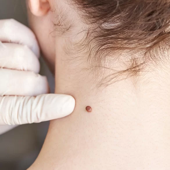
An atypical nevus (dysplastic nevus, English: dysplastic nevus) is an acquired pigmented skin lesion with irregular clinical and histopathological structure, showing features intermediate between a benign melanocytic nevus and melanoma. It is characterized by asymmetry, irregular borders, variegated coloring and usually a diameter exceeding 5 mm. Microscopic examination reveals disturbances in the architecture of melanocytes (pigment cells producing melanin) and features of dysplasia, that is, abnormal cell maturation. The presence of numerous atypical nevi increases the risk of developing melanoma, particularly in individuals with a positive family history and a fair skin phototype.
Atypical mole – what is it?
An atypical nevus is a variant of a melanocytic nevus with an increased potential for malignant transformation compared with a common nevus, but it is not melanoma itself. It most commonly appears during adolescence and early adulthood, less frequently in children.
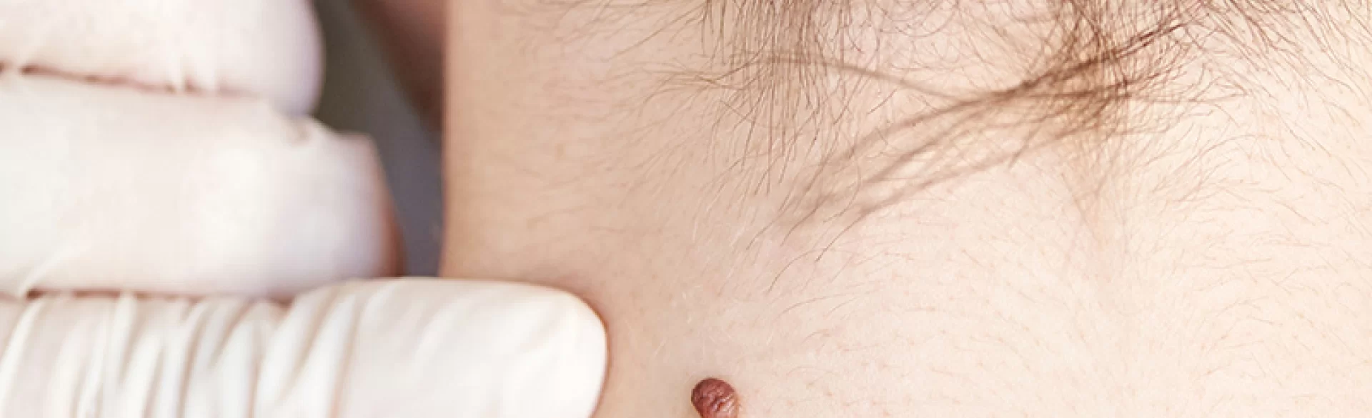
Clinically, these lesions meet some of the ABCDE criteria used to assess melanoma risk:
- A (asymmetry) – asymmetry of shape,
- B (border) – irregular, blurred borders,
- C (color) – non-uniform coloration (from light brown to dark brown, sometimes with a pinkish hue),
- D (diameter) – diameter usually > 5–6 mm,
- E (evolution) – change over time.
Histopathologically, the following are observed:
- irregular nests of melanocytes at the dermoepidermal junction,
- fibrosis of the dermis,
- features of cytologic dysplasia of varying degrees of severity (mild, moderate, or severe).
One distinguishes sporadic atypical nevi and the so-called dysplastic nevus syndrome (FAMMM – familial atypical multiple mole melanoma syndrome), in which numerous atypical nevi are present and there is an increased familial risk of melanoma.
Atypical mole and melanoma
Melanoma (Latin: melanoma malignum) is a malignant tumor originating from melanocytes, with a high metastatic potential. An atypical nevus is not a melanoma, but constitutes a marker of increased risk of its development.
The risk of transformation of a single atypical nevus into melanoma is relatively low, however the following are of significant importance:
- the number of atypical nevi (>5–10),
- a family history of melanoma,
- fair skin (phototype I–II),
- intense, intermittent exposure to UV radiation,
- sunburns in childhood.
In clinical practice the greatest challenge is differentiating an atypical nevus from an early melanoma. For this purpose the following are used:
- dermoscopy – a non-invasive examination that magnifies the image of the pigmented structure,
- videodermoscopy with digital archiving of lesions,
- histopathological examination after excision of the lesion.
In the case of clinical progression (enlargement, change of color, the appearance of itching, bleeding) an urgent dermatological consultation is necessary. Early diagnosis of melanoma radically improves the prognosis – in the stage limited to the epidermis (melanoma in situ) the cure rate exceeds 95%.
Atypical nevus – removal
Management depends on the clinical picture, the result of dermatoscopy, and the patient's risk factors. Not every atypical nevus requires removal – in many cases regular dermatological follow-up (every 6–12 months) and skin self-examination are sufficient.
Indications for surgical excision include:
- dynamic change in appearance,
- suspicion of melanoma,
- moderate or severe dysplasia on histopathological examination,
- difficulty monitoring the lesion (e.g., location on the back).
The standard is surgical excision of the lesion with a margin of healthy skin and mandatory histopathological examination. Destructive techniques (laser, electrocoagulation, cryotherapy) are not recommended in cases of suspected atypical nevus, because they prevent microscopic assessment of the entire lesion.
For prevention, the key measures are:
- photoprotection (SPF 30–50, year-round protection),
- avoidance of tanning beds,
- regular monitoring of nevi using dermatoscopy,
- education on skin self-examination.
In the case of multiple atypical nevi, ongoing dermatologic care and an individualized monitoring plan are recommended.

