Sutton's nevus
back to main page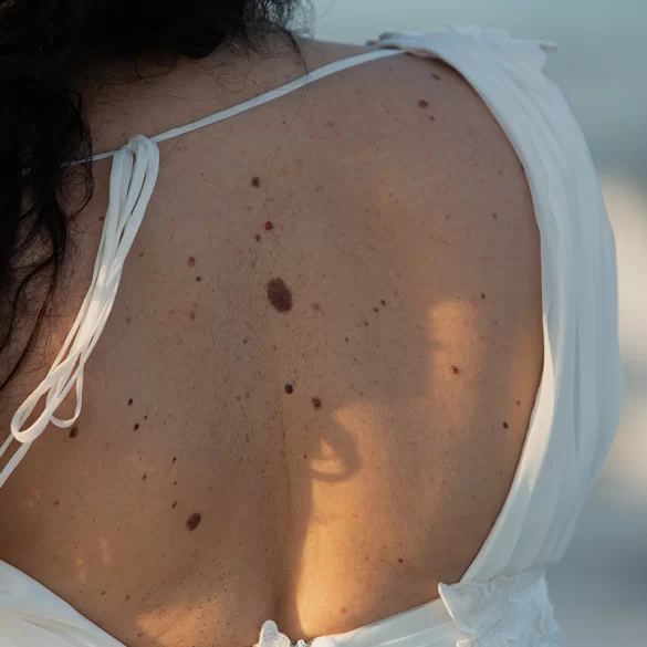
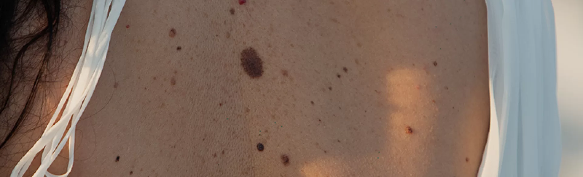
A Sutton's nevus (halo nevus) is a specific type of pigmented mole characterized by the presence of a light, depigmented border surrounding a central melanocytic nevus. This change is most commonly benign and occurs mainly in children and young adults, although it can appear at any age. The depigmented zone results from an immune reaction directed against melanocytes, the cells that produce melanin. In its natural course, a Sutton's nevus may gradually disappear, and skin depigmentation may undergo partial or complete repigmentation over time. Although in most cases the lesion does not pose an oncological threat, it requires proper dermatological diagnosis because in rare situations it may coexist with other pigmentation disorders or mask lesions of a neoplastic nature.
Sutton's Nevus – Causes
The etiology of Sutton's nevus is not fully clear, but the current state of knowledge indicates a key role of immunological mechanisms. The body recognizes melanocytes as antigens and triggers an autoimmune response, leading to their gradual destruction both within the nevus and in the surrounding skin.
- Autoimmune reactions – particularly associated with the activation of cytotoxic T lymphocytes.
- Genetic predisposition – more common in individuals with a family history of autoimmune diseases.
- Association with vitiligo – in some patients, Sutton's nevus coexists with vitiligo patches or precedes their appearance.
- UV radiation exposure – intense tanning can act as a trigger for the immune response.
- Hormonal factors and stress – presumably modulate the activity of the immune system.
It is important to emphasize that the occurrence of Sutton's nevus itself does not imply a systemic disease. In most patients, it is an isolated, benign skin lesion that does not require aggressive treatment but only dermatological observation.
Sutton's nevus – treatment methods
The therapeutic management of Sutton's nevus primarily depends on the clinical presentation, the patient's age, and the results of dermatoscopic examinations. In the vast majority of cases, active treatment is not implemented, and the foundation is regular observation.
- Dermatological control and dermatoscopy
Regular examinations allow for the assessment of the nevus's symmetry, color, borders, and any changes over time. - Photoprotection
Protection against UV radiation is crucial as skin lacking melanin is more susceptible to sun damage. - Conservative management
In the absence of atypical features, surgical treatment is not undertaken. - Supportive treatment of accompanying disorders
If Sutton's nevus coexists with vitiligo or other autoimmune diseases, treatment is directed at the underlying condition.
Pharmacological preparations are not routinely used to "treat" Sutton's nevus itself. Attempts to modulate the immune response are made only in the context of extensive pigmentation disorders, not a single nevus.
Sutton's nevus – removal
Removal of a Sutton's nevus is not standard practice and should only be considered in strictly defined situations. It is crucial to differentiate between the lesion and melanoma or atypical moles.
- ambiguous dermoscopic appearance,
- asymmetry, irregular borders, or changes in the color of the nevus,
- rapid enlargement of the lesion,
- the presence of subjective symptoms (itching, bleeding),
- significant oncological burden in the family history.
The most preferred method is surgical excision of the nevus with a margin of healthy skin and mandatory histopathological examination. Destructive methods (e.g., laser) are not recommended for pigmented lesions of unclear nature, as they prevent a full histological assessment.
The decision to remove should always be preceded by a dermatological consultation and based on medical, not solely aesthetic, considerations.
Treatments for Sutton's nevi
In the case of Sutton's nevi, "surgical treatment" is not discussed in the classical sense, however, modern dermatology and aesthetic medicine offer procedures that support diagnosis, observation, and improve skin quality in depigmented areas.
- Advanced digital dermoscopy – allowing for image archiving and comparing changes over time.
- Videodermoscopy – useful in monitoring patients with numerous nevi.
- Procedures improving the condition of the skin around the lesion – regenerative and biostimulating procedures, applied only to healthy skin, not covering the nevus itself.
- Interdisciplinary consultations – in cases of coexisting vitiligo or other autoimmune diseases.
It should be clearly emphasized: aesthetic procedures are not performed directly on Sutton's nevus unless it has been unequivocally classified as a benign lesion and has not been previously surgically removed. Oncological safety always takes precedence over aesthetic aspects.
