Loss of pigment
back to main page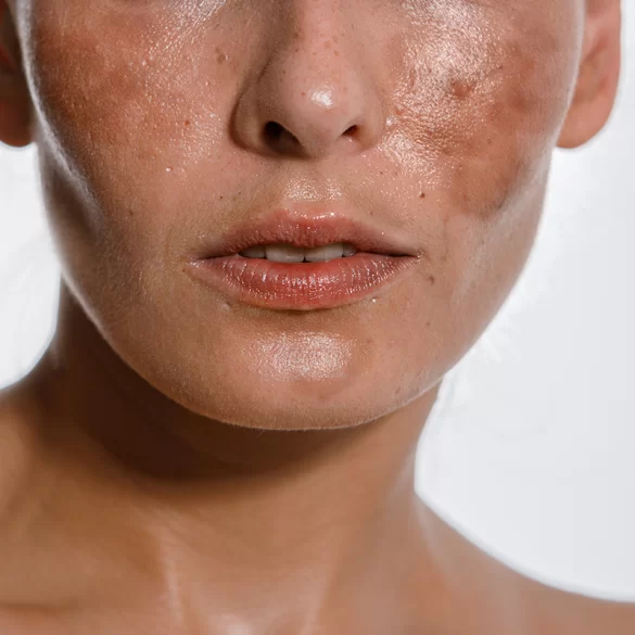
Loss of pigment (depigmentation) is a process involving the reduction or complete disappearance of melanin – a natural skin pigment produced by melanocytes. This phenomenon can be local or generalized and results from functional disorders of these cells, their damage, or immunological destruction. Depigmentation is a significant dermatological problem because it affects the uniformity of skin tone, and in some cases it may be a symptom of systemic diseases. It is most commonly observed in the course of vitiligo, post-inflammatory hypopigmentation, and post-traumatic discoloration.
Pigment loss – causes
The causes of pigment loss are complex and include both local and systemic factors. A key role is played by malfunctions of melanocytes and processes leading to their damage or elimination.
The most important mechanisms include:
- autoimmune factors – in vitiligo, melanocytes are destroyed by T lymphocytes (especially CD8+), and autoimmune diseases, such as thyroid diseases, often coexist,
- oxidative stress – an excess of reactive oxygen species leads to damage of pigment cells and disorders of melanogenesis,
- enzymatic disorders – abnormal activity of tyrosinase (a key enzyme for melanin synthesis) limits pigment production,
- post-inflammatory hypopigmentation – a consequence of skin inflammation (e.g., acne, atopic dermatitis, injuries),
- chemical and drug-induced factors – contact with toxic substances or certain medications can lead to depigmentation,
- genetic factors – congenital disorders of melanogenesis (e.g., albinism),
- mechanical and iatrogenic damage – e.g., after intensive dermatological procedures, burns, or chronic friction.
The so-called Koebner phenomenon, which consists of the appearance of lesions at the sites of skin injury, is also significant in the pathogenesis of depigmentation. This mechanism is observed particularly often in the course of vitiligo.
Pigment loss – most common areas
Loss of pigment can occur in various areas of the body, but there are locations particularly predisposed to the appearance of depigmentation changes. This results from both the distribution of melanocytes and exposure to environmental factors and micro-injuries.
The most commonly observed locations include:
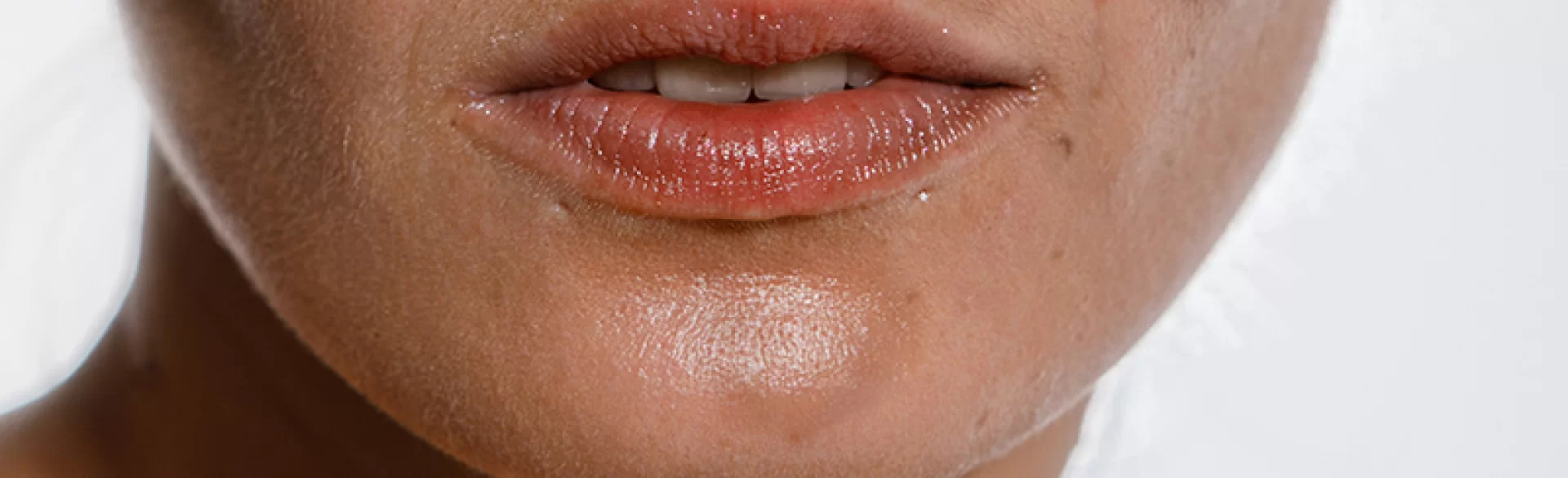
- face – especially the areas around the eyes and mouth, where the skin is thin and sensitive,
- hands and fingers – areas exposed to frequent contact with irritants,
- elbows and knees – areas of increased friction,
- genital area – due to the specific structure of the skin,
- scalp – depigmentation can also involve the hair (leukotrichia),
- trunk – especially in generalized forms of depigmenting diseases.
It is also of clinical significance that the location of the changes affects the effectiveness of therapy – well-vascularized areas, such as the face, usually show a better response to treatment than the limbs.
Loss of pigment – how to recover
The process of restoring pigmentation (repigmentation) depends on the cause of depigmentation, its duration, and the preserved activity of melanocytes. Treatment is multidirectional and includes both dermatological methods and procedures supporting skin regeneration.
1. Dermatological treatment
The basis consists of therapies aimed at modulating the immune response and stimulating melanogenesis:
- topical glucocorticosteroids – anti-inflammatory and immunosuppressive effect,
- calcineurin inhibitors (tacrolimus, pimecrolimus) – particularly effective in treating lesions on the face,
- UVB 311 nm phototherapy (NB-UVB) – stimulates melanocyte proliferation and pigment cell migration,
- PUVA therapy – used less frequently due to a higher risk of side effects.
2. Regenerative and procedural therapies
Modern aesthetic medicine uses methods supporting the skin's microenvironment and melanocyte activity:
- excimer laser therapy (308 nm) – selective stimulation of depigmentation foci,
- needle mesotherapy – delivery of active substances that improve cellular metabolism,
- platelet-rich plasma (PRP) – regenerative effect through growth factors,
- microneedling – induction of repair processes and improvement of skin blood supply.
3. Surgical methods
In cases resistant to treatment, grafting techniques are used:
- autologous melanocyte transplants,
- transplants of epidermis containing pigment cells.
4. Supportive management
The therapy is complemented by:
- photoprotection – reducing the contrast between healthy and depigmented skin,
- antioxidants – reduction of oxidative stress,
- psychological support – especially important in chronic diseases such as vitiligo.
Loss of pigment – prognosis
The prognosis in the case of pigment loss depends on the etiology, the duration of changes, and the location of depigmentation foci. In clinical practice, significant variability in the course of the disease is observed – from stable to progressive changes.
The most important factors affecting the prognosis include:
- cause of depigmentation – post-inflammatory hypopigmentation is often reversible, while vitiligo is chronic and recurrent,
- duration of changes – the earlier the treatment is implemented, the greater the chance of repigmentation,
- location – the face and neck respond better to therapy than the hands and feet,
- presence of active melanocytes – a necessary condition for effective repigmentation,
- patient's age and general health status.
In many cases, it is possible to achieve partial or significant repigmentation; however, this process is long-term and requires systematic therapy. Complete restoration of pigment is not always achievable, especially in advanced and long-lasting forms of depigmentation.
From a clinical point of view, it is important to realistically plan the therapy and individualize the treatment, taking into account both medical and psychological aspects.