Mole examination
back to main page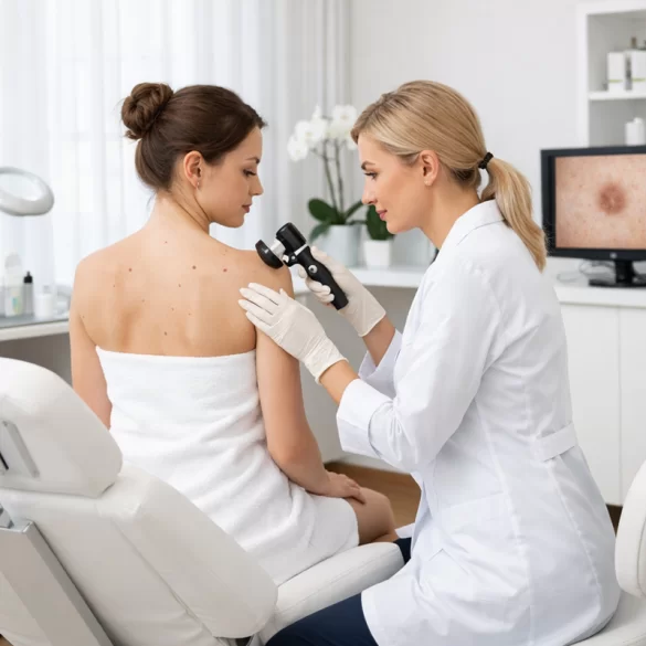
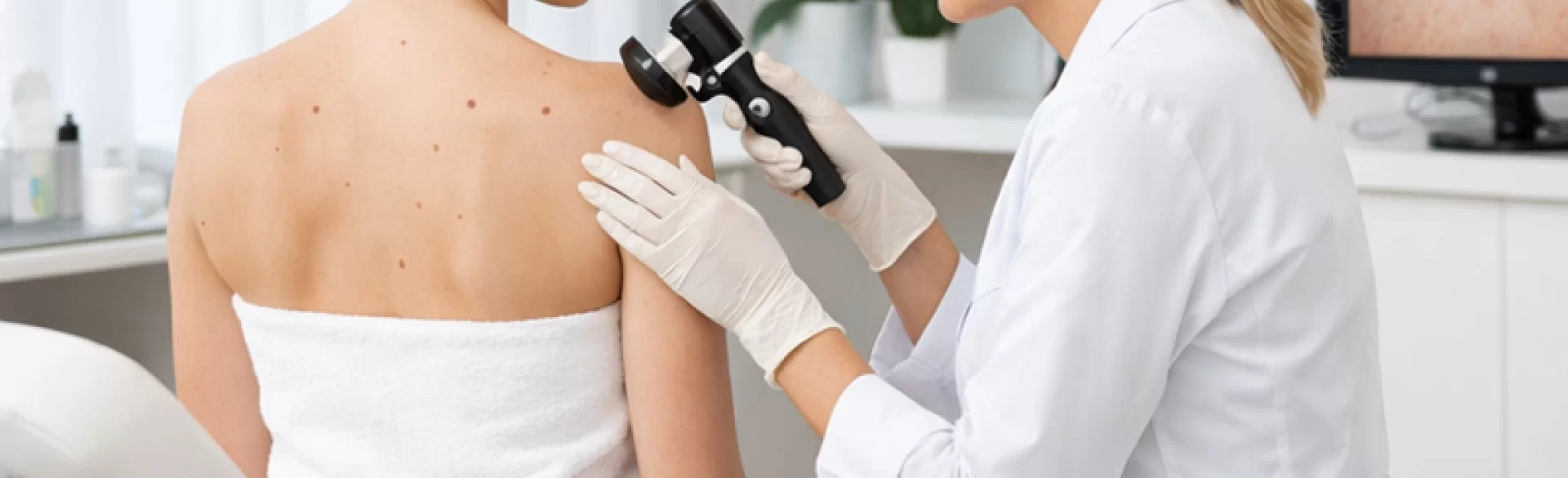
Mole examination is a specialized assessment of pigmented (melanocytic) lesions and other skin changes to rule out precancerous and cancerous skin conditions, particularly malignant melanoma. The diagnosis is based on a clinical examination of the entire skin and dermoscopy, a non-invasive method that allows for the evaluation of structures not visible to the naked eye. Regular monitoring of moles is a crucial element of cancer prevention, especially for individuals with a fair skin type, numerous moles, a family history of melanoma, and those with a history of intense UV exposure.
Mole examination under the National Health Fund (NFZ)
In Poland, mole examination can be performed under the National Health Fund, but the availability of the service depends on several organizational and formal factors.
Basic Information:
- A referral to a dermatology clinic is required (from a primary care physician).
- Waiting time depends on the region and the number of contracted services.
- The visit usually includes:
- medical interview,
- evaluation of moles in a physical examination,
- dermoscopy of selected lesions.
Under the National Health Fund, it is also possible to:
- refer for surgical removal of a suspicious lesion,
- histopathological examination of the removed mole (standard in oncological diagnostics).
Systemic Limitations:
- lack of possibility for a preventive "review of all moles" without clinical indications in some facilities,
- long waiting times,
- lack of access to digital videodermatoscopy (monitoring changes over time) in most public centers.
From the perspective of evidence-based medicine, it is crucial that any lesion exhibiting alarming features (asymmetry, irregular borders, color change, rapid growth, bleeding) is assessed urgently, regardless of the funding pathway.
Mole examination – how much does it cost
The cost of a private mole examination depends on the scope of diagnostics and the technology used.
Factors affecting the price:
- dermatological consultation (usually 200–400 PLN),
- manual dermoscopy included in the visit price or as an additional service,
- digital videodermatoscopy (with image archiving and comparison over time),
- the number of lesions assessed (some facilities limit the number of moles included in the basic price).
Approximate price ranges in Warsaw:
- dermatological consultation with dermoscopy: 250–450 PLN,
- mole mapping (so-called total body mapping): 400–900 PLN,
- surgical removal of a lesion: 500–1500 PLN (depending on location and size),
- histopathological examination: 150–400 PLN.
From a medical perspective, the key is not so much "how much it costs" but whether the examination includes dermoscopy and potential digital monitoring, which significantly increases the sensitivity of detecting melanoma at an early stage.
Mole examination – what it looks like
The examination of moles is a non-invasive, painless procedure that typically lasts from 15 to 30 minutes, depending on the number of lesions.
1. Medical Interview
The doctor assesses:
- the number and dynamics of skin lesions,
- the occurrence of melanoma in the family,
- history of sunburns,
- use of tanning beds,
- skin phototype (Fitzpatrick scale).
2. Examination of the entire skin surface
According to current dermatological recommendations, the assessment should include:
- the skin of the face and scalp,
- torso (front and back),
- limbs,
- areas of the hands, feet, and nails,
- intimate areas (if indicated).
This is important as melanoma can develop in areas not directly exposed to UV radiation.
3. Dermoscopy
A dermoscope is a magnifying device (10–20×) that allows for the assessment of:
- the pigment network,
- globular structures,
- architectural asymmetry,
- the presence of atypical structures (e.g., pseudopods, radial streaming).
Algorithms used in diagnostics include:
- ABCDE (Asymmetry, Border, Color, Diameter, Evolution),
- 7-point Glasgow scale,
- analysis of dermoscopic patterns.
In the case of suspicious lesions, the standard procedure is surgical excision of the lesion with a margin and histopathological examination, which remains the only method to confirm the diagnosis of melanoma.
Regular examination of moles (every 6–12 months for individuals at risk) is one of the most effective elements of secondary prevention of skin cancer. Early detection of melanoma is associated with over 90% 5-year survival, whereas advanced stage dramatically worsens the prognosis. In preventive medicine, vigilance is not overzealousness – it is the standard procedure.

