Pubic symphysis
back to main page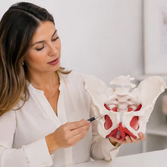

The pubic symphysis (Latin: symphysis pubica) is an anatomical structure connecting the two pubic bones in the anterior part of the pelvic girdle. It is a cartilaginous joint and consists of an interpubic disc made of fibrocartilage and the surrounding stabilizing ligaments. Despite its limited physiological mobility, the pubic symphysis plays a key role in pelvic biomechanics, enabling the transfer of loads between the trunk and the lower limbs. This structure undergoes particular changes during pregnancy and childbirth, when its flexibility increases, which can lead to pain and impaired stability.
Pubic symphysis – what is it?
The pubic symphysis is the central element of the anterior part of the pelvic ring, functionally cooperating with the sacroiliac joints. Its primary characteristic is the ability to absorb compressive and tensile forces that arise during everyday activities such as walking, standing, or changing body position.
The structure of the pubic symphysis includes:
- interpubic disc – a fibrocartilaginous structure with shock-absorbing properties,
- superior and inferior pubic ligaments – responsible for mechanical stability,
- surrounding soft tissues – including the fascia and muscles that form part of the central stabilization system.
Biomechanically, the pubic symphysis:
- contributes to the so-called pelvic ring, providing structural integrity,
- transmits loads between the lower limbs and the spine,
- works in conjunction with deep muscles (including the transverse abdominal muscle, the diaphragm, and the pelvic floor muscles),
- participates in the regulation of intra-abdominal pressure.
Under physiological conditions the mobility of the pubic symphysis is minimal (approx. 1–2 mm of displacement), which allows maintenance of the balance between stability and flexibility.
Pubic symphysis – pain during pregnancy
Pain of the pubic symphysis in pregnancy is a common clinical phenomenon and is part of a broader syndrome referred to as pelvic girdle pain (PGP – pelvic girdle pain). Its pathogenesis is primarily related to hormonal and biomechanical changes occurring in a woman’s body.
The hormone relaxin plays a key role, which causes:
- relaxation of the pelvic ligaments,
- increased tissue extensibility,
- reduced passive stability of joint structures.
Additionally, there is:
- a shift in the body’s center of gravity,
- increased lumbar lordosis,
- overloading of the anterior part of the pelvis.
Pain symptoms include:
- pain in the area of the pubic symphysis that worsens with walking,
- difficulties when standing up, turning over in bed, climbing stairs,
- a feeling of instability or “spreading” of the pelvis,
- radiation of pain to the groins, thighs, or sacral area.
In more advanced cases there may be a diastasis of the pubic symphysis (diastasis symphysis pubis), i.e., a pathological increase in the distance between the pubic bones above physiological values (usually >10 mm).
Pubic symphysis – causes of pain
Pubic symphysis pain is multifactorial and can occur both in pregnant women and in physically active individuals or after injuries. Disturbances in the balance between pelvic stability and mobility are of key importance.
The most common causes include:
- instability of the pelvic ring resulting from weakness of the deep muscles,
- dysfunction of the transversus abdominis and pelvic floor muscles,
- sacroiliac joint dysfunctions,
- overuse from asymmetric activities (e.g., running, unilateral sports),
- mechanical pelvic injuries,
- degenerative changes of cartilage and ligaments,
- fascial dysfunctions affecting force transmission.
An important element of the pathomechanism is dysfunction of the so-called central stabilization system (core). Under normal conditions:
- the diaphragm,
- the transversus abdominis,
- the pelvic floor muscles,
- and the paraspinal muscles
act synergistically to stabilize the pelvis and control intra-abdominal pressure. Their dysfunction leads to overload of the pubic symphysis and increased pain symptoms.
In clinical practice it is also crucial to differentiate pubic symphysis pain from other conditions, such as:
- sacroiliac joint pain,
- lumbar disc disease (discopathy),
- hip pathologies,
- overuse syndromes of the adductor muscles.
Pubic symphysis – when to see a specialist
A specialist consultation is indicated in situations when pubic symphysis pain affects daily functioning or shows a tendency to worsen. Early intervention helps reduce the risk of chronic pelvic instability.
Indications for consulting a specialist include:
- pain that impairs walking, standing, or changing position,
- persistent symptoms after childbirth,
- a feeling of pelvic instability,
- limited physical activity,
- suspected separation of the pubic symphysis,
- no improvement despite rest.
Diagnostics include:
- functional examination of the pelvis,
- clinical tests assessing stability,
- in selected cases imaging studies (ultrasound, X-ray).
Therapeutic management is multifaceted and includes:
- urogynecological physiotherapy aimed at restoring central stabilization,
- manual therapy (work on soft tissues and joints),
- individually tailored exercises activating the deep muscles,
- education on movement ergonomics and daily activities,
- use of pelvic stabilization belts.
In modern therapeutic approaches, methods supporting tissue regeneration and improving the quality of soft tissues are also used, such as:
- fascial therapy,
- neuromobilization techniques,
- physical modalities supporting reparative processes.
A comprehensive approach allows not only pain reduction but also the restoration of proper biomechanical pelvic function and the prevention of symptom recurrence.