Stretch marks on thighs
back to main page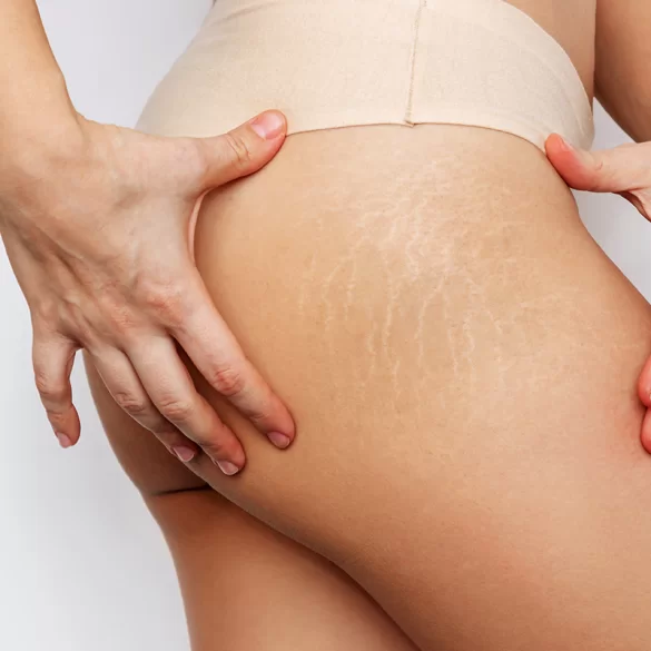
Stretch marks on the thighs (Latin striae distensae) are linear, spindle-shaped atrophic bands that form as a result of damage to the collagen and elastin fibers in the dermis. They represent a form of atrophic scarring and are the result of excessive stretching of the skin and hormonal disturbances affecting its structure. They are most commonly located on the lateral and medial surfaces of the thighs, and less frequently on the posterior area. This issue affects both women and men, although it is more frequently observed in women during puberty, pregnancy, and weight fluctuations. Stretch marks do not pose a health risk; however, due to their aesthetic and psychological impact, they are a common reason for dermatological and aesthetic consultations.
Stretch marks on thighs – what causes them
The pathogenesis of stretch marks is multifactorial. Mechanical stretching of the skin and hormonal changes affecting the metabolism of fibroblasts—cells responsible for the production of collagen and elastin—play a key role.
Key etiological factors:
- Sudden weight gain – stretching of the skin beyond its adaptive capacity.
- Puberty – rapid body growth and hormonal changes (estrogens, androgens).
- Pregnancy – the effect of steroid hormones and mechanical tension of the skin.
- Intensive strength training – rapid increase in muscle mass.
- Endocrine disorders – e.g., hypercortisolism, Cushing's syndrome.
- Prolonged glucocorticosteroid therapy (topical or systemic).
- Genetic predispositions – differences in the structure of the extracellular matrix.
At the histological level, the following observations are made:
- disorganization and fragmentation of type I and III collagen fibers,
- reduction in the number of elastin fibers,
- thinning of the epidermis,
- decreased fibroblast activity.
Contrary to popular belief, stretch marks are not merely an "aesthetic defect" resulting from weight gain. Their development is closely related to the skin's biological response to hormonal and mechanical factors.
Stretch marks on thighs – what do they look like
The clinical appearance of stretch marks depends on their stage of development. Two main phases are distinguished:
1. Inflammatory Phase –
striae rubrae
- pink, red, or purple color,
- delicate raised bands,
- possible itching or a feeling of skin tightness,
- presence of enlarged blood vessels.
This is an active stage where the inflammatory process and degradation of supporting fibers are most intense. It is also the moment when therapeutic intervention yields the best results.
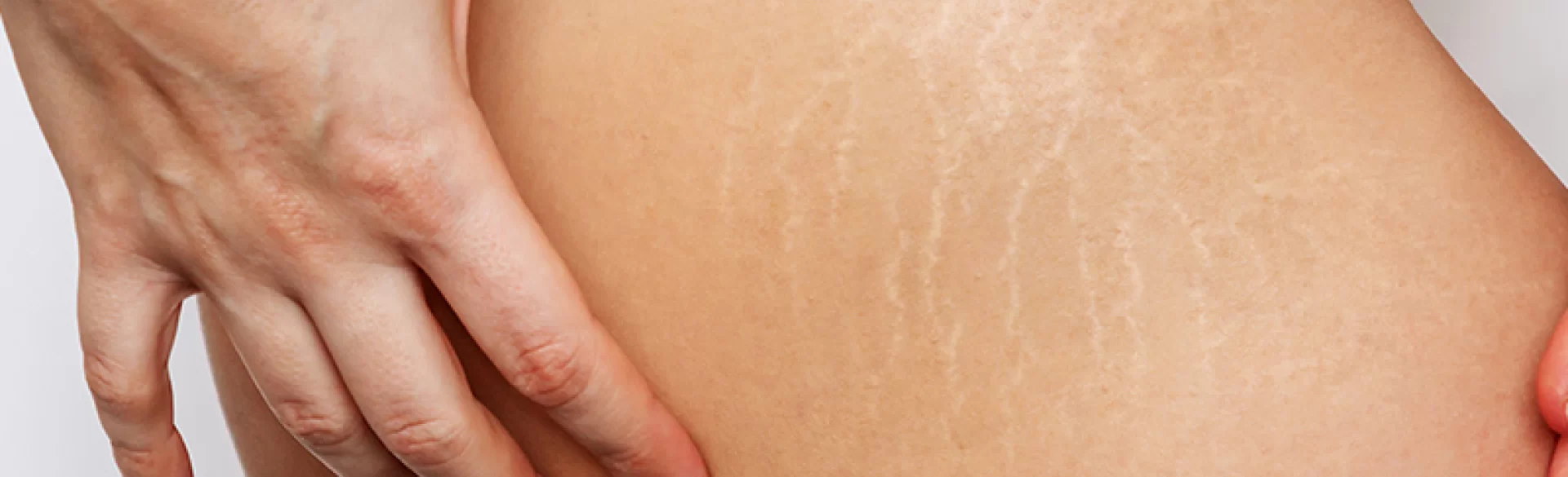
2. Atrophic Phase –
striae albae
- white, pearly, or silvery color,
- sunken, thin structure,
- lack of pigmentation (hypopigmentation),
- reduced skin elasticity within the lesion.
In this phase, stretch marks have the characteristics of a permanent atrophic scar. The skin within the lesion does not tan because the number of melanocytes is reduced.
Typical locations on the thighs include:
- lateral parts of the thighs (most common),
- medial surfaces,
- subgluteal areas.
Stretch marks usually align parallel to the lines of skin tension.
Stretch marks on thighs – how to remove
It should be clearly emphasized: completely removing stretch marks is not possible as they are a form of scarring. The goals of therapy are:
- to reduce and narrow the changes,
- to improve skin tone,
- to thicken the skin,
- to improve its elasticity.
The effectiveness of the therapy depends on:
- the age of the stretch marks,
- the depth of the changes,
- the type of skin,
- the individual's regenerative response.
Methods with documented effectiveness:
- Fractional laser therapy (ablative and non-ablative) – induction of controlled microdamage and neocollagenesis.
- Microneedle radiofrequency (RF) – simultaneous mechanical and thermal stimulation of the dermis.
- Carboxytherapy – improvement of microcirculation and stimulation of regeneration through CO₂ administration.
- Microneedling (micro-needle mesotherapy) – stimulation of fibroblasts.
- Chemical peels (e.g., TCA) – controlled exfoliation and skin remodeling.
- Combined therapies – enhancing effectiveness through multi-level action.
Topical preparations (retinoids, hyaluronic acid, vitamin C) can support therapy, but their effectiveness as monotherapy is limited.
It is crucial to start treatment early – red stretch marks respond much better than white ones.
Treatments for stretch marks on thighs
In the treatment of stretch marks on thighs, procedures are used to stimulate controlled skin remodeling and induce the synthesis of new collagen.
At the Beauty Embassy in Warsaw, the following are used:
- Microneedle Radiofrequency (DeAge EX) – 3D RF technology allowing spatial energy delivery and deep remodeling of the dermis.
- Medical Carboxytherapy – improves oxygenation and microcirculation in the affected area.
- Medical Microdermabrasion – supports therapy in the initial phase.
- TCA Medical Peels – stimulate the regeneration of the epidermis and dermis.
- Combined RF + Mesotherapy Treatments – increase the intensity of neocollagenesis.
In selected cases, it is possible to combine therapy with:
- stimulating treatments (e.g., platelet-rich plasma mesotherapy),
- skin firming treatments (e.g., monopolar radiofrequency).
Treatment usually includes a series of 3–6 procedures at intervals of 4–6 weeks. The collagen remodeling process lasts up to 6 months after completing the therapy.
The effects of therapy include:
- reduction in the visibility of stretch marks,
- improvement in skin texture,
- increased tightness and elasticity,
- evening out the skin tone.
The choice of method should be preceded by a dermatological qualification and an assessment of the phase of changes.

