Subcutaneous tissue fibrosis
back to main page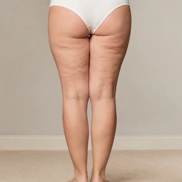

Subcutaneous tissue fibrosis means the excessive accumulation and abnormal remodeling of extracellular matrix components, primarily collagen fibers, in the layer located beneath the dermis. The tissue then becomes less elastic, harder, and less mobile relative to the underlying structures. The change may be local, for example after an injury, surgery, liposuction, injection, or inflammation, or it may be an element of fibrous cellulite or a systemic disease. The term "fibrosis" itself describes a tissue process, not a single specific disease. Diagnosis should take into account medical history, palpation, and the appearance of the skin, and in ambiguous cases, also ultrasonography. Sudden hardening, pain, redness, or a rapidly growing lump require medical evaluation.
Subcutaneous tissue fibrosis - what is it
The subcutaneous tissue, also called the hypodermis, consists of adipocytes (fat cells), vessels, nerves, and connective tissue septa connecting the dermis to the deeper fascia. Its extracellular matrix undergoes constant physiological remodeling. In the process of fibrosis, this balance shifts toward excessive synthesis and insufficient degradation of collagen.
Activated fibroblasts and myofibroblasts are of key importance. Under the influence of mediators, especially transforming growth factor beta (TGF-β), they produce collagen, fibronectin, and other structural proteins. If the damaging or inflammatory stimulus persists for too long, normal repair can turn into permanent scarring.
Fibrosis may involve:
- fibrous septa between fat lobules;
- tissue surrounding a scar, implant, or site of repeated injections;
- an area after surgery, radiotherapy, trauma, or chronic inflammation;
- subcutaneous tissue in the course of certain diseases, including systemic sclerosis, morphea, eosinophilic fasciitis, or lipodermatosclerosis;
- thickened connective tissue septa contributing to the dimples characteristic of cellulite.
Not every hardening under the skin is fibrosis. A lipoma, epidermoid cyst, hematoma, fat necrosis, calcification, superficial thrombophlebitis, abscess, or neoplastic tumor can feel similar.
Subcutaneous tissue fibrosis - how to feel it under your fingers
On palpation, fibrotic tissue may feel harder, dense, fibrous, or "adherent" to deeper layers. The skin-subcutaneous fold may be thicker and harder to lift, and shifting the skin relative to the fascia is limited. Sometimes, bands, irregular thickenings, or small, poorly demarcated nodules can be felt. Pressure or stretching may cause tenderness, but painless fibrosis is also possible.
Self-examination may include comparing symmetrical areas of the body:
- assessing the softness and elasticity of the tissue;
- gently lifting the skin fold;
- shifting the skin in several directions;
- checking for the presence of bands, indentations, or restricted mobility;
- observing pain, swelling, and changes in skin color and temperature.
Palpation does not reliably confirm the type of lesion. If the induration is new, unilateral, enlarging, well-demarcated, or persists over time, medical consultation is advised. High-frequency ultrasonography can visualize the architecture of subcutaneous tissue, septal thickness, edema, and the presence of focal lesions. In selected cases, elastography, magnetic resonance imaging, laboratory tests, or a biopsy are used.
Urgent evaluation is required for: rapidly increasing pain, redness, marked warmth, fever, ulceration, significant unilateral limb swelling, or a hard, immobile mass.
Subcutaneous tissue fibrosis - why does it occur
Fibrosis is the end result of an abnormally prolonged repair response. Cellular damage activates platelets, macrophages, and other immune cells. The released cytokines stimulate fibroblasts, which transform into contractile myofibroblasts and increase the production of the extracellular matrix. TGF-β is one of the main regulators of this process. Chronic hypoxia, oxidative stress, mechanical disturbances, and an imbalance between matrix metalloproteinases and their inhibitors are also significant.
The most common groups of causes include:
- trauma and surgical procedures, including liposuction, body contouring surgery, and complicated wound healing;
- repeated injections, local hematomas, fat necrosis, or a foreign body reaction;
- chronic inflammation or infection of subcutaneous tissue;
- radiotherapy, which can lead to progressive vascular damage and fibrosis;
- chronic venous insufficiency and venous hypertension, especially in lipodermatosclerosis of the lower legs;
- autoimmune and inflammatory diseases, in which induration may involve the skin, subcutaneous tissue, and fascia;
- cellulite with an established structure, associated, among other things, with the structure of fibrous septa, mechanical tension, adipose tissue distribution, and skin quality.
Obesity can promote adipose tissue fibrosis through adipocyte hypertrophy, local hypoxia, macrophage infiltration, and chronic low-grade inflammation. However, it is not a necessary condition for the occurrence of fibrosis or cellulite.
Subcutaneous tissue fibrosis - how does it differ from cellulite
Fibrosis is a histopathological process, whereas cellulite, scientifically referred to as gynoid lipodystrophy, is a characteristic alteration in skin topography, most commonly affecting the thighs and buttocks. It manifests as dimples, waviness, and an "orange peel" appearance. Fibrosis may contribute to its development, but these terms are not synonymous.
Feature | Subcutaneous tissue fibrosis | Cellulite |
Nature of the change | Excessive deposition and remodeling of the extracellular matrix | Multifactorial unevenness of the skin surface |
Clinical presentation | Induration, bands, thickening, restricted mobility | Dimples, bulges, and waviness of the skin |
Location | Any area, depending on the cause | Most commonly thighs, buttocks, hips, and abdomen |
Sex dependence | Occurs in women and men | Affects mainly post-pubertal women |
Causes | Trauma, surgery, inflammation, radiotherapy, systemic diseases | Fibrous septa anatomy, skin properties, adipose tissue, and hormonal factors |
Medical significance | May be a scar or a symptom of a disease | Usually an aesthetic condition, not an infection |
Anatomical and imaging studies indicate that cellulite depressions are primarily associated with connective tissue septa anchoring the skin. Between them, adipose tissue bulges the skin surface. Thickening or shortening of specific septa can perpetuate the depressions, but cellulite does not mean a generalized "overgrowth" of the entire subcutaneous tissue. It should also not be confused with bacterial infection of connective tissue, referred to in the English-language literature as cellulitis.
Subcutaneous tissue fibrosis - what treatments loosen it
The management depends on the cause. Postoperative, post-inflammatory, and systemic disease-related fibrosis require different treatment than the septa contributing to cellulite. Before aesthetic therapy, active inflammation, thrombosis, decompensated venous insufficiency, neoplastic lesions, and contraindications specific to a given technology must be ruled out.
The Ambasada Urody offer includes the following methods that may be applicable in the treatment of dense tissue, fibrous cellulite, or coexisting edema:
- shockwave therapy, including LipoShock and SkinShock: acoustic pulses induce mechanical stress, improve local blood circulation, and can support the remodeling of collagen septa; among non-invasive methods, acoustic wave therapy has some of the better, although still limited, data regarding cellulite;
- Icoone® Med: multi-micro alveolar vacuum massage mobilizes the skin-subcutaneous fold, supports drainage, and improves tissue mobility;
- LPG Endermologie: mechanical vacuum-roller massage can reduce stiffness, swelling, and the visibility of irregularities;
- VelaShape® and Body Mix: combine mechanical action with thermal energy or other modules, supporting surface smoothing and skin remodeling;
- radiofrequency, INDIBA® 448 kHz, and Accent Prime: controlled tissue heating primarily affects skin collagen, its tension, and quality; the effect on deep, persistent fibrous bands may be limited;
- Onda: microwaves primarily target localized adipose tissue and contour improvement, which is why the method is mainly relevant when fibrosis or cellulite is accompanied by an excess of adipocytes;
- carboxytherapy: intradermal or subcutaneous administration of carbon dioxide may temporarily increase blood flow and support tissue remodeling, but the quality of clinical evidence is limited;
- medical pressotherapy: reduces fluid stasis and swelling, but does not remove mature scar tissue on its own;
- lipolytic mesotherapy: primarily affects localized adipose tissue rather than fibrosis directly, which is why it requires strict medical qualification.
Non-invasive methods can improve tissue compliance, mobility, and appearance, but they do not guarantee the complete removal of mature fibrosis. In the case of individual, deep cellulite septa, a longer-lasting effect may be provided by severing the fibrous bands, i.e., subcision. However, this is not a universal treatment for every induration and must be performed by a properly trained physician. Therapy should be tailored to the diagnosis, not solely to the result of a palpation examination.

