Vascular changes
back to main page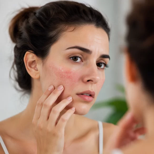
Vascular lesions are abnormalities within the blood vessels of the skin and subcutaneous tissue, resulting from their dilation, damage, proliferation, or structural disorders. They may be congenital or acquired and cover a wide spectrum of entities – from mild telangiectasias to complex vascular malformations. These lesions are of significant clinical importance for both aesthetic and health reasons, as they can accompany systemic diseases, hormonal disorders, or venous insufficiency. Their diagnosis and treatment require an interdisciplinary approach and consideration of the etiology, depth, and dynamics of the lesions.
Vascular lesions – types
Classification of vascular lesions is based on their structure, formation mechanism, and the nature of blood flow. In clinical practice, several main groups are distinguished:
1. Superficial vascular lesions (telangiectasias and erythema)
The most commonly encountered lesions include:
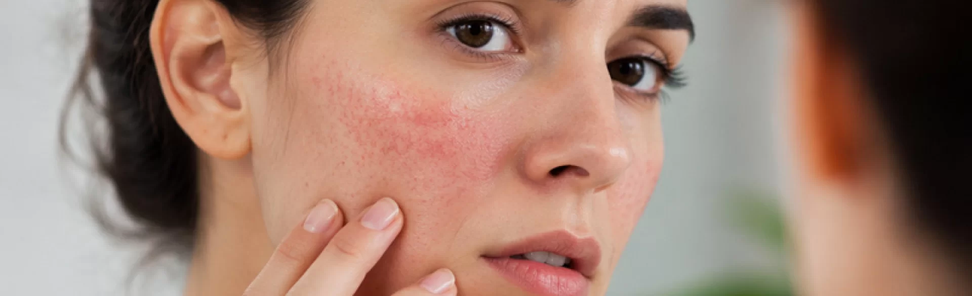
- telangiectasias (so-called „spider veins”) – permanent dilations of small capillaries, visible as thin, red or bluish-red lines,
- vascular erythema – chronic skin redness resulting from vascular hyperreactivity,
- erythrosis – persistent erythema, often associated with vascular-prone skin or rosacea.
The mechanism behind these changes is related to the loss of elasticity in vessel walls and the disruption of their tone regulation.
2. Hemangiomas
These are benign vascular tumors resulting from the proliferation of endothelial cells:
- capillary hemangiomas – frequently occur in infants and may resolve spontaneously,
- cavernous hemangiomas – located deeper, with a higher potential for complications.
Hemangiomas are neoplastic (benign) in nature, which distinguishes them from malformations.
3. Vascular malformations
These are congenital developmental defects of the vessels, present from birth and growing proportionally with the body:
- capillary malformations (e.g., „port-wine stain” spots),
- venous malformations,
- arteriovenous malformations (AVM) – characterized by an abnormal connection between arteries and veins.
These lesions do not undergo spontaneous regression and often require specialized treatment.
4. Lesions associated with venous insufficiency
This group includes:
- varicose veins of the lower limbs,
- venectasias and reticular vein dilations,
- chronic venous insufficiency, leading to discoloration and ulcerations.
Their pathogenesis is linked to the impairment of venous valve function and an increase in venous pressure.
5. Traumatic and inflammatory lesions
- petechiae and bruises (ecchymosis) – the result of vascular damage,
- purpura – associated with coagulation disorders,
- vascular lesions secondary to inflammatory states or autoimmune diseases.
6. Hormonal and environmental changes
- vascular spider veins during pregnancy,
- dilated vessels resulting from UV exposure,
- changes related to menopause or hormonal therapy.
Classification Summary
Lesion type | Nature | Dynamics |
|---|---|---|
Telangiectasias | acquired | progressive |
Hemangiomas | benign tumors | often regression |
Malformations | congenital | constant |
Varicose veins | acquired | progressive |
Purpura/petechiae | secondary | transient |
Vascular lesions – treatment
Therapeutic management of vascular lesions depends on their type, depth, location, and accompanying symptoms. Modern medicine utilizes both conservative methods and advanced procedural technologies.
1. Conservative and pharmacological treatment
In the case of mild lesions and as supportive therapy, the following are used:
- phlebotropic drugs (e.g., diosmin, hesperidin) – improve vascular wall tone,
- vessel-sealing preparations (vitamin C, rutin),
- anti-inflammatory treatment – especially in the course of rosacea,
- compression therapy – in venous insufficiency (compression stockings).
Lifestyle modification also plays a significant role:
- avoiding exposure to high temperatures,
- sun protection (UV filters),
- reduction of factors exacerbating erythema (alcohol, spicy foods).
2. Laser therapy and light technologies
One of the most effective methods for treating vascular lesions is the use of light energy:
- vascular laser (e.g., Nd:YAG, KTP) – selective photothermolysis of hemoglobin leads to vessel closure,
- IPL (Intense Pulsed Light) – broadband light reducing erythema and fine telangiectasias.
The mechanism of action involves the absorption of energy by hemoglobin and controlled damage to the vessel wall without affecting surrounding tissues.
3. Sclerotherapy
A method used mainly in the treatment of:
- telangiectasias of the lower limbs,
- varicose veins and venectasias.
It consists of injecting an obliterating substance (e.g., polidocanol) into the vessel lumen, which causes:
- endothelial damage,
- vessel closure,
- its gradual resorption.
4. Electrocoagulation and radiosurgery
Techniques using electrical energy:
- electrocoagulation – protein coagulation and closure of small vessels,
- radiosurgery – precise removal of lesions with minimal tissue damage.
They are used especially in the treatment of individual punctate lesions.
5. Surgical treatment
Indicated in the case of:
- large vascular malformations,
- advanced varicose veins,
- lesions with complications (e.g., bleeding, ulcerations).
Includes:
- classic surgical removal,
- endovascular procedures (e.g., embolization).
6. Modern supportive methods
In clinical practice, therapies improving microcirculation and skin condition are also used:
- fractional laser therapy – skin remodeling and improvement of its quality,
- LED light therapies – anti-inflammatory and strengthening effect,
- treatments stimulating microcirculation (e.g., vacuum massages).
7. Choice of therapy – deciding factors
The selection of the treatment method should take into account:
- lesion type (hemangioma vs telangiectasia),
- vessel depth and diameter,
- location (face vs limbs),
- presence of concomitant diseases,
- patient expectations.
Combined therapy is often used, which allows for optimal clinical effects.