Drooping nasal tip
back to main page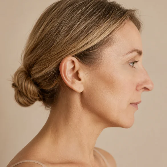
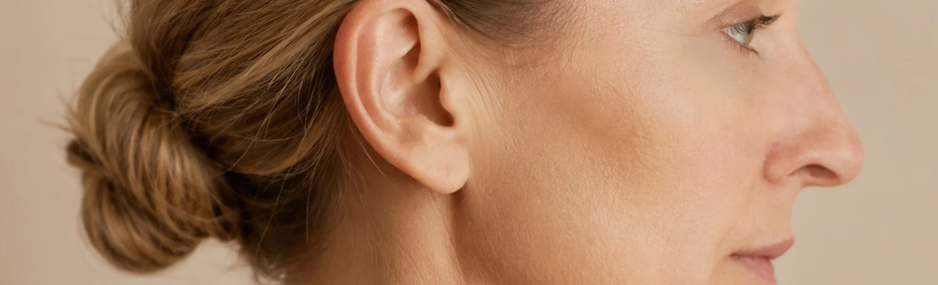
A drooping nasal tip, medically referred to as nasal tip ptosis, indicates its excessive downward rotation relative to the nasal dorsum, nasal base, and upper lip. It can be a congenital trait, the result of trauma or surgery, as well as an element of the physiological aging of the face. Its appearance depends on the structure of the alar cartilages, nasal septum, supporting ligaments, skin, and the activity of facial muscles. In some people, the drooping occurs mainly while smiling, while in others, it is also visible at rest. It is not a disease in itself, but significant ptosis can coexist with impaired nasal patency, especially when accompanied by weakness of the nasal valve. Therefore, the choice of correction method requires an assessment of both aesthetics and the respiratory function of the nose.
Drooping tip of the nose - why the nose droops with age
The nose does not grow throughout life in the same way it does during the developmental period. Its apparent lengthening and drooping result primarily from the gradual remodeling of cartilage, bone, and soft tissues. Nasal aging is a multilayered process, and tip ptosis rarely has just a single cause.
The most important mechanisms include:
- weakening of the connections between the nasal cartilages, including the ligaments and fibrous attachments stabilizing the alar cartilages;
- flattening or downward deflection of the greater alar cartilages, which form the primary framework of the tip;
- reduced cartilage elasticity, associated with changes in the composition of the extracellular matrix and its progressive calcification;
- weakened support of the nasal septum, the columella, and the anterior nasal spine of the maxilla;
- bone resorption of the midface, especially around the pyriform aperture and the maxilla, which alters the support of the nasal base;
- the action of the depressor septi nasi muscle, which can chronically pull the tip downward;
- changes in the skin and subcutaneous tissue, including loss of elasticity, the effects of gravity, and, in some individuals, skin thickening and increased mass of the tip tissues.
As a result of these processes, derotation occurs—meaning a downward rotation of the tip—along with, in some cases, a reduction in its stable projection beyond the facial plane. The nose may appear longer, and a previously minor dorsal convexity becomes more prominent. The nasolabial angle also decreases.
The severity of these changes depends on baseline anatomy, skin thickness, sex, age, past injuries, and prior nasal surgeries. Patients who have undergone previous rhinoplasty require particular evaluation, as cutting or weakening the natural support mechanisms can lead to secondary tip ptosis. If the drooping is accompanied by breathing difficulties, a sensation of alar collapse, or chronic obstruction, an ENT or surgical consultation is necessary.
Drooping nose tip - why is it more visible when smiling
Drooping of the nasal tip during a smile is dynamic in nature. This means that its severity depends not only on the structure of the cartilages, but also on the action of the muscles of the nose and upper lip. A key role is played by the depressor septi nasi muscle (musculus depressor septi nasi). Its fibers connect the area of the maxilla and the orbicularis oris muscle with the mobile part of the septum and the columella.
During a broad smile, the following can occur simultaneously:
- pulling of the columella and nasal tip downwards by the depressor septi;
- elevation of the upper lip;
- elevation of the alar base by the levator labii superioris alaeque nasi muscle;
- widening of the nasal base and increased visibility of the nostrils;
- reduction of the nasolabial angle and optical elongation of the nose.
The effect is particularly pronounced when the depressor septi muscle is strong or hyperactive, and the cartilaginous support elements of the tip are weak. Drooping during a smile can also occur in young individuals without ptosis visible at rest. Therefore, it should not be automatically considered a sign of aging.
A proper examination includes an assessment of the face and profile at rest, during a gentle smile, and at maximum smile. A photograph taken only in a neutral position may not reveal the dynamic deformity. The physician also evaluates the movement of the upper lip, the width of the nasal base, the position of the alae, as well as changes in tip projection and rotation.
Distinguishing the dynamic form from the static one has therapeutic significance. When muscle hyperactivity is dominant, its action can be temporarily limited with botulinum toxin. If the cause is insufficient cartilaginous support, reducing muscle activity alone may yield a minor or short-lived effect. In such cases, methods to improve tissue support or surgical treatment are considered.
Drooping nasal tip - how to assess the nasolabial angle
The nasolabial angle is one of the parameters used in nasal profile analysis. It is formed around the subnasale point, which is the junction of the columella and the upper lip. In practice, several methods are used to determine it, which is why results from different methods are not always directly comparable.
In classic photographic assessment, the angle between the following is analyzed:
- the line describing the inferior border of the columella or the axis of the nostril;
- the line running along the anterior surface of the upper lip.
The measurement should be performed on a standardized profile photograph, with a neutral head position, relaxed lips, and the natural position of the jaws maintained. Photographs at rest and during a maximum smile should be analyzed separately. A decrease in the angle value when smiling indicates dynamic downward rotation of the tip.
Frequently cited reference ranges are approximately 90-100° in men and 95-110° in women, but they are neither a diagnostic standard nor a universal aesthetic model. Aesthetic preference studies have pointed to even narrower ranges, but their results depended on the population, photograph preparation method, and evaluators. The angle should be interpreted in conjunction with:
- nasal projection and length;
- the position of the upper lip and chin;
- nostril exposure;
- columellar shape;
- skin thickness;
- sex, ethnicity, and individual facial proportions.
Self-measurement using an app may carry a significant perspective error. Selfie-type photos, especially those taken from a close distance with a wide-angle lens, distort nasal projection. The nasolabial angle should also not be the sole qualification criterion for a procedure. Professional evaluation includes palpation of tip support, analysis of the septum, nasal patency, and the relationship between the nose, lips, and chin.
Drooping nose tip - how to lift it without surgery
Non-surgical correction can improve the appearance of mild or moderate ptosis, but it does not remodel cartilage and does not replace rhinoplasty in the case of significant deformity or breathing disorders. The method is selected based on the dominant mechanism.
Botulinum toxin type A
The product can be administered into the depressor septi nasi muscle to temporarily reduce its activity. The method is most justified when the tip droops primarily while smiling. The effect consists of limiting the dynamic downward pulling of the tip, rather than a mechanical lifting of the entire nasal structure. The result is temporary and usually develops within a few days.
Possible side effects include smile asymmetry, a temporary change in upper lip mobility, and an insufficient or excessive effect. Botulinum toxin will not correct a significant lack of cartilaginous support. The botulinum toxin nasal tip lift procedure is part of the range of treatments offered by Ambasada Urody.
Nose reshaping with hyaluronic acid
Cross-linked hyaluronic acid can be precisely administered into selected areas of the nasal dorsum, columella, or base of the nose. Proper placement of the product allows for increasing tip support, softening an apparent hump, and visually improving the profile. However, this is an additive procedure: it does not reduce the nose, but adds volume at strategic points.
The nose is among the injection areas with increased vascular risk. Accidental injection of filler into an artery can cause ischemia and skin necrosis, and, in rare cases, visual disturbances or loss of vision. The procedure requires an excellent knowledge of anatomy, asepsis, cautious dosing, and the immediate availability of hyaluronidase. Nose modeling with hyaluronic acid is a procedure available at Ambasada Urody.
Aptos lifting threads
Barbed threads can mechanically support the tip tissues and improve the nasolabial angle. The secondary biostimulatory effect is associated with a healing response and collagen remodeling around the threads. The method may be considered for selected, mild deformities, but its predictability and evidence base are lower than in the case of surgical cartilage stabilization.
Complications include hematomas, infection, asymmetry, palpability or visibility of the threads, skin dimpling, material displacement, and unsatisfactory durability of the correction. Aptos threads are available at Ambasada Urody and are also used for minimally invasive nose reshaping.
If the ptosis is significant, structural in nature, or coexists with nasal obstruction, rhinoplasty remains the most predictable method. It may include strengthening the tip support with cartilage grafts, septum stabilization, correction of the alar cartilages, and, in appropriate cases, modification of the depressor septi muscle. The final qualification should be based on a medical examination, and not solely on the value of the nasolabial angle.

