Facial retaining ligaments
back to main page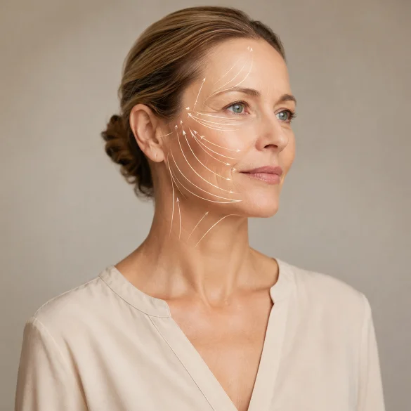

Facial retaining ligaments, referred to in the literature as facial retaining ligaments, are bands and septa of fibrous connective tissue that stabilize the skin, adipose tissue, and the superficial musculoaponeurotic system of the face, or SMAS, relative to deeper-lying fascia or periosteum. They form a three-dimensional network of anchoring points and boundaries between soft tissue compartments. Thanks to it, individual facial regions maintain their proper position while simultaneously being able to perform the movements necessary during facial expression, chewing, and speaking. Ligaments do not act alone. The appearance of the face is determined by the cooperation of bones, muscles, fascia, fat compartments, and skin. Their anatomy is of fundamental importance in plastic surgery and in planning procedures that contour the cheeks, facial oval, and jawline.
Facial retaining ligaments - what are they
Facial retaining ligaments are structures composed mainly of dense connective tissue containing collagen fibers. They run transversely to the facial layers, connecting superficial structures with deep ones. They should not be equated with articular ligaments. Their role is not to stabilize a joint, but to maintain a specific position of soft tissues and control the extent of their displacement.
In surgical anatomy, two basic types of these structures are distinguished:
- osteocutaneous ligaments, originating from the periosteum and passing through subsequent layers towards the skin, e.g., zygomatic and mandibular ligaments;
- fasciocutaneous ligaments, which originate within the deep fascia and connect it to the SMAS and subcutaneous tissue, e.g., parotid-cutaneous ligaments and part of the masseteric ligaments.
The SMAS is a continuous fibromuscular layer located between the subcutaneous tissue and the deep structures of the face. Ligaments pass through the SMAS or connect with it, creating a mechanical system that:
- transfers forces generated by mimetic muscles;
- limits excessive displacement of the skin and fat pads;
- separates adjacent fat compartments;
- delineates the boundaries of anatomical gliding spaces;
- influences the location of grooves, folds, and characteristic contours of the facial surface.
Blood vessels and branches of the facial nerve often run in the vicinity of the ligaments. For this reason, they serve as important landmarks, but also as high-risk areas during surgery and injections. Procedures performed near these structures require a very good knowledge of the layered anatomy of the face.
The term “ligament” is sometimes used inconsistently. Some structures take the form of distinct, dense bands, while others resemble a diffuse system of fibrous septa. Differences in nomenclature also stem from individual variation and differing methods of anatomical dissection.
Facial retaining ligaments - where are they located
The ligamentous system encompasses all tiers of the face. Individual ligaments differ in their attachment site, course, and functional significance.
Structure | Approximate location | Main anatomical significance |
|---|---|---|
Temporal ligament and areas of temporal adhesions | lateral part of the forehead and temple | stabilization of tissues in the temporofrontal region |
Orbicularis retaining ligament | along the inferior and lateral orbital rim | attachment of the orbicularis oculi muscle to the periosteum, defining the boundaries of the infraorbital region |
Zygomatic ligaments | over the zygomatic prominence | strong anchoring of the cheek and support for the tissues of the midface |
Maxillary ligaments | medial part of the cheek, maxillary region | stabilization of tissues adjacent to the nasolabial fold |
Masseteric ligaments | along the anterior border of the masseter muscle | connection of the masseteric fascia to the SMAS and skin, defining the mobile zone of the cheek |
Parotid-cutaneous ligaments | over the parotid gland | stabilization of the lateral, relatively immobile part of the face |
Mandibular ligament | anterior part of the mandibular body, near the labiomandibular fold | anchoring of the tissues of the lower face and defining the anterior border of jowling |
Between the ligaments, there are gliding spaces, where tissue layers can move relative to each other. The facial system therefore does not resemble a uniform sheet sagging under the influence of gravity. It is a mosaic of strongly attached and more mobile areas. The boundaries of these areas influence the formation sites of nasolabial folds, labiomandibular grooves, and cheek sagging colloquially referred to as “jowls”.
Of particular importance is the so-called line of ligaments, which encompasses the lateral orbital adhesions, zygomatic, parotid-masseteric, and mandibular ligaments. It separates the relatively stable tissues of the lateral part of the face from the more mobile tissues of the anterior cheek. This concept is useful in facelift surgery, although the actual architecture of fibrous connections is three-dimensional and more complex than a single line.
Facial retaining ligaments - how they change with age
Ligament aging cannot be considered in isolation from other layers. With age, simultaneous changes occur in the craniofacial skeleton, fat compartments, muscles, SMAS, fasciae, and skin. Their rate varies individually and depends, among other factors, on anatomy, sex, exposure to ultraviolet radiation, tobacco smoking, hormonal changes, and body weight fluctuations.
The traditional model of aging assumes a gradual stretching and weakening of ligaments, leading to the gravitational displacement of tissues. However, contemporary anatomical studies indicate that the mechanism is more complex. The deep portions of some ligaments, especially at their bony and fascial attachments, remain relatively strong. In contrast, changes may affect:
- superficial branches passing through the SMAS and subcutaneous tissue;
- septa separating fat compartments;
- tissues located between fixed anchoring points;
- the bony base to which the ligaments attach;
- the quality of collagen in the skin, fasciae, and supporting structures.
In some regions, the ligament continues to firmly hold the tissues, while an adjacent, more mobile compartment bulges or descends. A visible boundary then forms between the fixed and displaced area. Examples include the nasolabial fold, the labiomandibular fold, and the anterior border of the "jowl".
The picture of aging is simultaneously influenced by:
- bone remodeling, including changes in the projection of the maxilla, orbit, chin, and mandible;
- loss or redistribution of fat volume, where some compartments atrophy and others may increase in volume;
- deterioration of skin quality, resulting from the degradation of collagen, elastin, and the extracellular matrix;
- changes in muscle tone, particularly of the platysma, depressor anguli oris, and masseter muscles;
- repetitive facial movements, which affect the tissues adjacent to the points of fixed attachment.
Therefore, the statement that ligaments simply "stretch and sag" is an oversimplification. The observed clinical effect results from the spatial remodeling of the entire facial support system.
Facial retaining ligaments - why they determine the jawline
A distinct jawline is created through the interplay of bone shape, chin projection, fat distribution, tension of the skin and platysma muscle, and ligamentous anchoring points. The most important fibrous structure of this area is the mandibular ligament, located in the anterior part of the mandibular body, adjacent to the labiomandibular sulcus.
The mandibular ligament supports the tissues of the anterior part of the lower face. Laterally located tissues are more mobile. When they lose volumetric support, undergo remodeling, or become lax, they can bulge over the inferior border of the mandible. The ligament then acts as a fixed anterior boundary of the developing "jowl". At the same time, volume loss in front of the ligament can create a depression referred to as the prejowl sulcus. The contrast between the depression and the bulge further disrupts the smoothness of the contour.
Loss of jawline definition is also influenced by:
- decreased chin projection or bone changes of the mandible;
- excess adipose tissue in the submental area;
- volume loss in the lateral cheek;
- laxity of the skin and subcutaneous tissue;
- activity of platysmal bands;
- masseter muscle hypertrophy;
- descent of the tissues of the mid and lower face.
Non-surgical treatments do not "rebuild" ligaments nor do they release them in a surgical sense. However, they can act on other elements of the system responsible for the contour. In the offer of Ambasada Urody, procedures used for the loss of facial oval and an undefined jawline include:
- facial modeling with hyaluronic acid, volumetric lifting, V-lifting, and Texas lift: replenish support in selected compartments, correct the prejowl sulcus, and increase the projection of the chin, mandibular angle, and mandibular body;
- calcium hydroxyapatite and poly-L-lactic acid: exert a biostimulatory effect by increasing collagen synthesis and improving the mechanical properties of tissues, though they do not replace the ligament;
- lifting threads: provide temporary mechanical repositioning of tissues and a secondary fibrotic response, but are not an equivalent of surgical SMAS suspension;
- HIFU SonoQueen: creates thermal coagulation points at a selected depth, initiating collagen remodeling and moderate tissue tightening;
- microneedle radiofrequency and monopolar or bipolar radiofrequency: controlled heating of the dermis and subcutaneous tissue leads to collagen remodeling and improved firmness;
- INDIBA: utilizes capacitive-resistive diathermy for controlled heating and supporting tissue remodeling;
- ablative and non-ablative fractional lasers: primarily improve the quality, density, and elasticity of the skin;
- endermologie and Icoone: act mechanically on the skin and subcutaneous tissue, supporting their mobilization and tension;
- Nefertiti Lift with botulinum toxin: weakens the excessive activity of the platysma muscle and selected depressor muscles, limiting their downward pull on the lower face.
The choice of method should stem from diagnosing the dominant cause. Adding filler will not correct significant excess skin, and intensive fat reduction can worsen laxity in a person with volume deficiency. In cases of advanced ptosis, surgical facelift remains the most effective method, with appropriate work on the SMAS and, depending on the technique, controlled release of selected ligaments. The mandibular region contains important vessels and the marginal mandibular branch of the facial nerve, which is why every injectable or surgical procedure requires medical qualification and a detailed knowledge of anatomy.

