Pigmented nevus
back to main page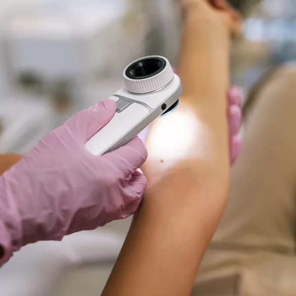

Pigmented nevus (Latin: naevus pigmentosus) is a localized skin lesion that arises from a focal accumulation of melanocytes — the cells that produce melanin, the skin pigment. Nevi may be congenital or acquired and can occur in various locations on the body. They most often appear as spots, papules, or slightly elevated lesions ranging in color from light brown to black. Most pigmented nevi are benign and do not cause symptoms, however some require dermatological monitoring due to the possibility of malignant transformation. Evaluation of nevi is an important element of melanoma prevention — one of the most aggressive malignant skin cancers.
Pigmented nevus – types
Pigmented nevi are a varied group of skin lesions that are classified based on histological structure, time of onset, and the location of pigment cells in the skin. Most commonly, acquired and congenital nevi are distinguished.
Acquired melanocytic nevi usually appear in childhood or adolescence and are the result of proliferation of melanocytes within the epidermis or dermis. Among them are, among others:
- Junctional nevi – melanocytes are located at the junction of the epidermis and dermis; lesions usually appear as flat, dark macules,
- Compound nevi – melanocytes are present both in the epidermis and the dermis; they are often slightly raised,
- Intradermal nevi – pigment cells are located mainly in the dermis; the lesions often take the form of dome-shaped papules.
Congenital nevi are present at birth or appear in the first months of life. They can reach various sizes – from small lesions to so-called giant nevi. Large congenital nevi are associated with a higher risk of melanoma development and require regular dermatological monitoring.
In dermatology, other variants of nevi are also distinguished, e.g.:
- Dysplastic (atypical) nevi – with irregular shape and varied pigmentation,
- Blue nevi – arising due to deep localization of melanocytes in the skin,
- Spitz nevi – characteristic lesions occurring more frequently in children and adolescents.
Accurate classification has clinical significance, because some types of nevi may show a greater tendency for malignant transformation.
Pigmented lesion – what does it mean
The presence of pigmented moles on the skin is a physiological phenomenon and affects the majority of the population. The number of moles in an adult can range from a few to even several dozen lesions. Their formation is influenced primarily by genetic factors and exposure to ultraviolet radiation.
The most important factors that favor the formation of moles include:
- genetic predispositions,
- exposure to UV radiation, especially in childhood,
- skin phototype – people with fair skin usually have more moles,
- hormonal changes (e.g., puberty or pregnancy).
In most cases pigmented moles are benign lesions and do not pose a threat to health. However, regular monitoring is important. Dermatologists recommend using the so-called ABCDE rule, which helps assess potentially concerning features of a lesion:
- A – asymmetry (asymmetry),
- B – irregular borders (border),
- C – varied color (color),
- D – diameter above 6 mm (diameter),
- E – evolution of the lesion (evolution), i.e., changes in shape, color, or size.
The appearance of such features may suggest malignant transformation and requires urgent dermatological consultation. Diagnostic methods include, among others, dermatoscopy, which allows assessment of the lesion's structure at high magnification.
Pigmented nevus – removal
Removal of pigmented nevi is performed for various indications: diagnostic, preventive, or aesthetic. The most important medical indication is suspicion of a neoplastic lesion or its atypical character on dermatoscopic examination.
The most commonly used methods for removing nevi include:
1. Surgical excision of the lesion
This method is considered the gold standard for nevi suspected of being neoplastic. The procedure involves removing the lesion along with a margin of healthy skin, and then sending the tissue for histopathological examination. This allows an unequivocal determination of the nature of the lesion.
2. Laser removal of nevi
In the case of lesions with a confirmed benign character, it is possible to use laser technologies that cause selective destruction of cells containing melanin. This method is used mainly for aesthetic purposes, e.g., for small pigmented lesions.
3. Electrocoagulation methods and dermatologic surgery
In some cases, electrocoagulation or other dermatologic techniques are used to remove minor skin lesions.
In clinical practice, the qualification of the lesion before the procedure is of particular importance. Not every pigmented lesion should be removed by laser or cosmetic methods. If melanoma is suspected, surgical excision and histopathological evaluation are necessary.
Regular dermatological check-ups and dermatoscopic examination of nevi are a basic element of skin cancer prevention. People with numerous nevi are also advised to perform periodic photographic documentation of the skin, which facilitates monitoring any changes in their appearance.