Scars from stitches
back to main page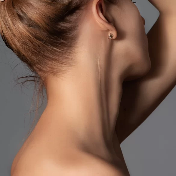
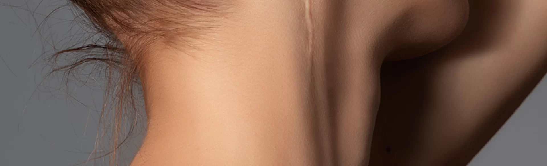
Suture scars are the result of surgical wound closure using surgical sutures, staples, or other techniques for approximating the edges of the skin. They represent the final stage of the physiological healing process, during which tissue continuity is restored by forming new connective tissue rich in collagen. The character, visibility, and quality of a scar depend on many factors: the type of wound, the suturing technique, skin tension, genetic predisposition, and the course of healing. A scar may take the form of a thin, pale line, but also a broad, hypertrophic thickening. Understanding the mechanisms of its formation enables the implementation of preventive and therapeutic measures that minimize its visibility.
Scars after stitches — when do they remain?
The formation of a scar is an inevitable consequence of damage to the dermis (dermis). If a wound involves only the epidermis, regeneration may occur without a permanent mark. However, in the case of wounds requiring sutures, deeper layers are damaged, resulting in the formation of scar tissue.
The healing process proceeds in three phases:
- Inflammatory phase (0–5 days) – activation of inflammatory cells and cleansing of the wound.
- Proliferative phase (up to 3–4 weeks) – proliferation of fibroblasts and intensive synthesis of type III collagen.
- Remodeling phase (up to 12–18 months) – replacement of type III collagen with type I collagen and reorganization of fibers.
The final appearance of a scar is assessed only after completion of the remodeling phase. Factors that increase the risk of hypertrophic scars or keloids are:
- location on the sternum, shoulders, back,
- excessive skin tension,
- wound infection,
- prolonged inflammation,
- genetic predisposition.
Suture scars "remain" whenever the injury involved the dermis, but their visibility can be greatly reduced through appropriate care and treatment.
Scars after stitches — what to apply
Topical management is key in the prevention of pathological scarring. Preparations are used only after complete wound healing and suture removal, when the epidermis is closed.
The best-documented efficacy is shown by:
- Medical silicones (silicone gels and sheets) – reduce transepidermal water loss (TEWL), regulate fibroblast activity, and limit scar hypertrophy.
- Preparations containing heparin, allantoin, and onion extract (Allium cepa) – may support collagen remodeling, although their efficacy is moderate.
- Ointments with vitamin A or E – there is no clear scientific evidence that they are superior to silicone.
Important elements of adjunctive therapy:
- sunscreen protection SPF 50+ for at least 6–12 months,
- gentle scar massage to improve tissue elasticity,
- avoiding stretching of the wound area in the first weeks.
In cases with a tendency toward hypertrophic scarring, early implementation of pressure therapy or injections of glucocorticosteroids is considered.
Suture scars – laser removal
Laser therapy is currently one of the best-documented methods for improving the quality of postoperative and post-traumatic scars. It should be emphasized that the goal of therapy is not to "excise" or completely remove the scar, but controlled remodeling of pathological collagenous tissue and improvement of its structure, color and elasticity. The mechanism of action is based on the phenomenon of selective photothermolysis – light energy of a specific wavelength is absorbed by water or hemoglobin, leading to microthermal damage and activation of skin repair processes.
In clinical practice the following are used primarily:
- Fractional CO₂ laser (ablative) – creates microcolumns of ablation within the scar, initiating intense neocollagenesis (formation of new type I collagen) and reorganization of fibers. Effective in hypertrophic and thickened scars.
- Er:YAG laser – acts more superficially, with a shorter recovery period; used mainly for normotrophic scars and minor surface irregularities.
- Pulsed dye laser (PDL, 585–595 nm) – reduces erythema and excessive vascularity of early scars by selectively closing pathological vessels.
The timing of therapy initiation is crucial. Increasing evidence shows that early implementation of laser therapy (as early as 4–8 weeks after wound healing and suture removal) can limit the development of hypertrophic scarring by modulating the inflammatory response and affecting fibroblast activity. In the case of mature scars, therapy also brings improvement, but usually requires a greater number of sessions.
The standard protocol includes:
- 3–6 treatments,
- intervals of 4–8 weeks,
- individual adjustment of parameters to skin phototype and scar location.
Clinical effects include:
- reduction in scar thickness and firmness,
- improved elasticity,
- reduction of redness,
- smoothing of the skin surface.
In selected cases therapy is combined with other methods, such as intralesional corticosteroid injections for hypertrophic scars, platelet-rich plasma (PRP), microneedle radiofrequency or microneedling, which enhances tissue remodeling efficacy.
It should be remembered that treatment effectiveness depends on scar type (normotrophic, hypertrophic, keloid), its age, location and the individual response of the patient. Laser therapy is a medical procedure requiring physician qualification and appropriate parameter selection, particularly in patients with darker skin phototypes who are at risk of postinflammatory hyperpigmentation.
From a clinical perspective, properly planned laser therapy is currently one of the most rational tools for treating suture scars, because it acts directly on the biological mechanisms of their remodeling rather than merely masking the aesthetic problem.

