Keratosis pilaris
back to main page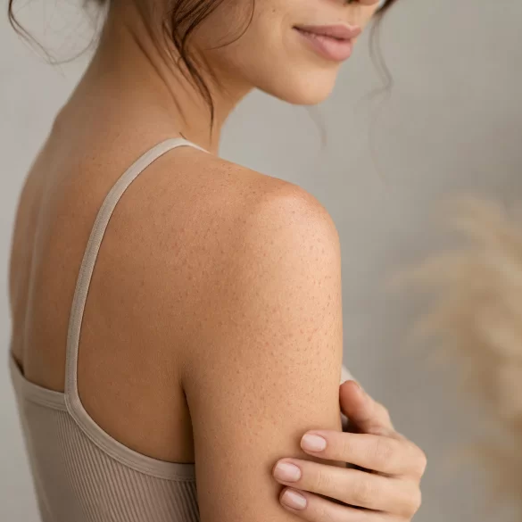
Follicular keratosis (keratosis pilaris) is a chronic, non-inflammatory disorder of epidermal keratinization, characterized by the excessive accumulation of keratin within hair follicles. As a result, small, rough bumps resembling “goosebumps” are formed. The lesions are cosmetic in nature, but in some patients they may coexist with dry skin, pruritus, or inflammation. The condition is very common, especially in children, adolescents, and individuals with atopy. Keratosis pilaris does not pose a threat to health, but it can significantly affect psychological comfort and self-esteem, especially with extensive severity of the lesions.
Keratosis pilaris - what is goose skin
Keratosis pilaris, colloquially referred to as “goosebumps,” is a disorder of the epidermal exfoliation process. Under normal conditions, cells of the stratum corneum are gradually renewed and shed from the skin surface. In the case of keratosis pilaris, there is an overproduction of keratin – a protein that builds the epidermis, hair, and nails. Keratin accumulates in the openings of hair follicles, leading to their clogging.
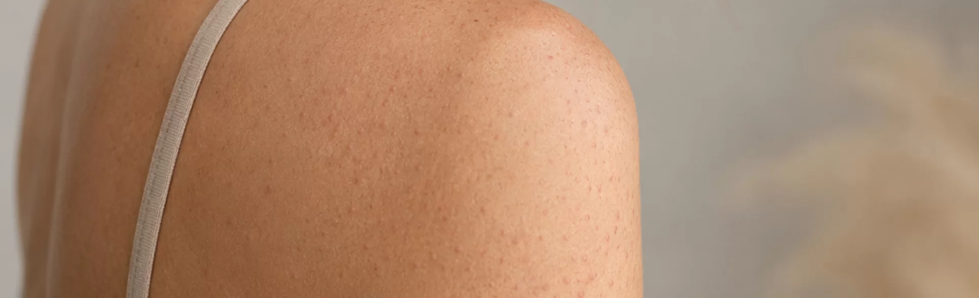
The lesions take the form of small perifollicular papules, often more palpable to the touch than visible to the eye. The skin becomes rough, uneven, and dry. In some patients, erythema appears around the papules, resulting from chronic micro-inflammation.
Keratosis pilaris is classified as a genodermatosis, i.e., a skin disease with a genetic basis. The condition often runs in families and is associated with atopic dermatitis, asthma, and allergies. Its characteristic course includes periodic flare-ups and remissions, with the severity of symptoms usually increasing in autumn and winter, when air humidity drops.
Although keratosis pilaris is not an infectious disease and does not lead to serious complications, it requires proper dermatological care. Improper treatment of the skin, intense rubbing, or aggressive scrubs can lead to worsening of the lesions and secondary inflammation.
Keratosis pilaris - symptoms and appearance
The most characteristic symptom of keratosis pilaris is small bumps located around the hair follicles. The lesions are usually skin-colored, white, pink, or red. To the touch, they resemble fine sandpaper or “tiny bumps.”
Typical symptoms include:
- skin roughness,
- small follicular bumps,
- dryness of the epidermis,
- redness around the hair follicles,
- a feeling of skin tightness,
- periodic itching,
- uneven skin texture.
In more severe cases, inflammatory lesions, post-inflammatory discoloration, or small scars resulting from scratching and chronic irritation may appear. In people with fair complexions, red dots dominate, whereas in patients with darker skin, brown discoloration is more often visible.
Symptoms usually worsen:
- in winter,
- at low air humidity,
- after hot baths,
- when using irritating cosmetics,
- in the course of atopy and severe skin dryness.
Keratosis pilaris can occur in various clinical variants. The most common form affects the arms and thighs, however, there are also rarer variants with extensive erythema or facial involvement. The chronic persistence of lesions with a tendency for periodic flare-ups remains characteristic.
In the differential diagnosis, atopic dermatitis, folliculitis, ichthyosis, and papular acne should be considered, among others. The diagnosis is usually established based on the clinical presentation without the need for additional tests.
Keratosis pilaris - causes and risk factors
The direct cause of keratosis pilaris remains an impaired process of keratinization, which is the maturation and shedding of epidermal cells. A key role is played by the excessive accumulation of keratin within the openings of hair follicles.
Genetic predisposition is of significant importance. In many patients, the condition runs in families, indicating a hereditary tendency to abnormal keratinization of the epidermis.
The most important risk factors include:
- atopic dermatitis,
- inhalation and food allergies,
- bronchial asthma,
- very dry skin,
- young age,
- disruptions of the hydrolipid barrier,
- low air humidity,
- frequent overheating of the skin,
- irritating skincare.
Keratosis pilaris is more commonly observed in children and adolescents. In some people, the symptoms decrease with age, but the condition can also persist into adulthood.
Modern research indicates that an abnormal epidermal barrier function may also play a role. Damage to the hydrolipid layer leads to increased transepidermal water loss (TEWL – transepidermal water loss), which exacerbates dryness and disrupts proper skin exfoliation.
Some people try to remove the lesions mechanically through intensive scrubs or skin brushing. Such actions often lead to the worsening of micro-inflammation and secondary redness. In therapy, regular, long-term skincare based on rebuilding the epidermal barrier and gentle keratolytic action is of significantly greater importance.
Keratosis pilaris - where it most commonly occurs
Changes characteristic of keratosis pilaris appear primarily in areas rich in hair follicles. The most common location is the lateral surfaces of the arms and the anterolateral parts of the thighs.
Typical areas of occurrence include:
- arms,
- thighs,
- buttocks,
- calves,
- cheeks,
- back.
In children, involvement of the face, especially the cheeks, is often observed. In such cases, the skin takes on a chronically red and rough appearance. In adults, changes on the arms and thighs predominate.
Less frequently, keratosis pilaris may affect:
- forearms,
- shoulder areas,
- abdomen,
- hairy skin.
The changes are usually symmetrical. During periods of exacerbation, the skin may become redder and noticeably rough. The severity of symptoms often increases after exposure to dry air, air conditioning, or improper skin care.
In the therapy of keratosis pilaris, the following are primarily used:
- urea preparations,
- lactic acid,
- salicylic acid,
- retinoids,
- exfoliating treatments,
- procedures improving hydration and rebuilding the skin barrier.
Modern cosmetology and aesthetic dermatology also utilize professional medical peels, laser treatments that reduce erythema, and highly skin-regenerating procedures. The offer of Ambasada Urody Clinic & SPA includes, among others, chemical peels, moisturizing and regenerating treatments, and procedures supporting the reconstruction of the skin's hydrolipid barrier.

